Unusual Presentation of Pneumoperitoneum Caused by Adenocarcinoma of the Pancreas Penetrating the Descending Colon Associated with Splenic Abscess
Takeshi Nishimura, Atsunori Nakao, Yasuaki Tsuchida, Ikuo Matsuda, Seiichi Hirota, Isamu Yamada and Joji Kotani
| Takeshi Nishimura1, Atsunori Nakao1*, Yasuaki Tsuchida2, Ikuo Matsuda2, Seiichi Hirota2, Isamu Yamada1 and Joji Kotani1 |
| Corresponding Author: Atsunori Nakao,MD, PhD, 1-1 Mukogawa, Nishinomiya, Hyogo 663-8501, Japan, Tel: +81-798-45-6514, Fax: +81-798-45-6813 E-mail: atsunorinakao@aol.com |
| Received: October 31, 2015 Accepted: December 09, 2015 Published: December 15, 2015 |
| Related article at Pubmed, Scholar Google |
Abstract
Background: Although pancreatic cancer is frequently locally aggressive, initial presentation as pneumoperitoneum is atypical. We report a case of ruptured spleen probably due to direct invasion of pancreatic cancer to descending colon presenting as peritonitis and pneumoperitoneum.
Methods: We examined the case of a woman who presented to our emergency department with acute abdominal pain associated with intra-splenic gas lesion.
Results: A 67-year-old woman was admitted to our department after three weeks of fever and left back pain. After discovering a splenic abscess via computed tomography (CT), antibiotics were administered and surgery was planned. On post-admission day four, she presented sudden diffuse abdominal pain, displaying muscle guarding and rebound tenderness. Abdominal CT demonstrated a pneumoperitoneum and intra-abdominal fluid around the spleen. A splenectomy, left hemicolectomy, lymph node dissection, and transverse colostomy were performed. The resected specimen presented a fistula from spleen to descending colon and perforation of splenic abscess. Histopathological and immunohistochemical examination demonstrated that pancreatic adenocarcinoma had infiltrated to the colon. The patient discharged on 22 days after surgery, and multiple metastases with liver and sacral bone was diagnosed after 36 days from discharge.
Conclusion: Since intra-splenic gas lesion following with pneumoperitonitis can be caused by many disorders, prompt recognition and early diagnosis help clinicians determine an appropriate management strategy.
Keywords |
||||||||
| Pneumoperitomeum; Pancreatic cancer; Splenic abscess | ||||||||
Introduction |
||||||||
| Splenic abscess is an extremely uncommon cause of pneumoperitoneum, particularly in patients without diabetes or compromised immune systems [1]. We encountered a case of splenic gas-containing rupture, causing acute abdomen and pneumoperitoneum. As far as we know, only seven cases of splenic abscesses presenting with pneumoperitoneum have been published in English literature [2-8]. Our patient was later diagnosed as pancreatic cancer, which locally invaded the spleen and descending colon, resulting in a gas-containing splenic abscess and pneumoperitoneum. Our report suggests that splenic gas associating to splenic abscess, which may be derived from pancreatic cancer, should also be expected when differentially diagnosing peritonitis or pneumoperitoneum, especially in patients with histories of upper left abdomen and lower left chest localizing signs and fever. | ||||||||
Case Presentation |
||||||||
| A 67-year-old woman complaining of left flank pain, bloody feces, and persistent high-grade fever lasting approximately three weeks was transferred to our hospital’s emergency department. | ||||||||
| Her family history was insignificant and her past medical history included uterine prolapse, cholecystectomy, and diabetes mellitus. | ||||||||
| On arrival, her blood pressure and heart rate were 112/62 mmHg and 122 beats per minute, respectively. Her abdomen was soft without rebound tenderness. Her laboratory test results on admission were as follows: white blood cell count, 10130/μl; red blood cell count, 329×104/μl; hemoglobin, 11.0 g/dl; platelet count, 15.3×10⁴/μl; C-reactive protein, 14.3 mg/dl; albumin, 3.3 g/dl; total bilirubin, 1.5 g/dl; D-dimer, 4.93 μg/ml; prothrombin time, 14.0 seconds; and carcinoembryonic antigen, 8.4 ng/ml, carbohydrate antigen 19-9, 9934 U/ml. | ||||||||
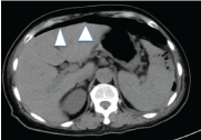
| Chest X-ray indicated clear lung field without cardiomegaly. Abdominal computed tomography (CT) disclosed a 12×8×13 cm nodular lesion containing air in the swollen spleen, as well as a thickening wall of the descending colon next to the spleen and inflammatory tissues around the tail of the pancreas. Therefore, fistula from splenic hilum to descending colon was suspected. Thrombus in splenic vein and gas containing lesion in spleen were visualized (Figure 1). A mass in the pancreas tail was not noticed initially. Echocardiography indicated no evidence of infective vegetation. We diagnosed the patient with splenic abscess due to colon cancer penetration, administered antibiotics (tazobactam/ piperacillin), and planned surgery after colonoscopy. | ||||||||
| On post-admission day four, the patient developed sudden chills and diffuse abdominal pain, displaying muscle guarding and rebound tenderness. Abdominal CT demonstrated a pneumoperitoneum and intraabdominal fluid around the spleen (Figure 2). The diagnosis of diffuse peritonitis due to colon perforation associated with splenic abscess was made and an emergency laparotomy was performed. Tight adhesion and inflammatory tissue were seen around hilum of spleen. Intraoperative findings demonstrated a fistula from descending colon to spleen. The patient underwent splenectomy, left hemi-colectomy, lymph node dissection, and colostomy of the transverse colon. No events occurred postoperatively and the patient was discharged 22 days later. | ||||||||
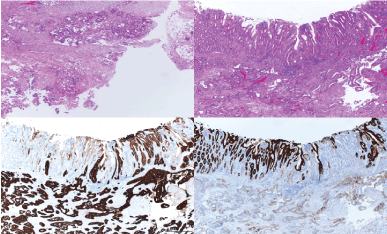
| The surgical resected specimen presented a fistula from descending colon to gas-containing lesion in the spleen and perforation of its tip probably due to splenic abscess. Although malignant cells were not identified in the spleen, histopathological examination demonstrated that adenocarcinoma was seen in the colon perforation site. The tumor cells were immunohistochemically positive for cytokeratin 7, and almost negative for cytokeratin 20, which is not typical for colon-derived cancer, but compatible with that of pancreatic cancer (Figure 3). | ||||||||
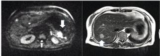
| After discharge on 22 days post operation, follow-up contrastenhanced magnetic resonance imaging showed a high signal intensity tumor at the tail of pancreas on diffusion weighted imaging 36 days after the operation, with multiple liver and sacral bone metastases (Figure 4). Therefore, according to these findings, we overall diagnosed the patient with splenic abscess due to invasion of pancreatic cancer to the colon. Chemotherapy and radical therapy were under consideration. | ||||||||
Discussion |
||||||||
| Intra-splenic gas is very infrequent, found as splenic abscess in 0.14-0.7% of autopsy cases, and often misdiagnosed because of non-specific symptoms [9]. Clinical manifestations for diagnosis of splenic abscess include left upper quadrant pain, tender mass, and fever [10]. However, only about 40% of cases present local signs. About 80% of patients present radiologic signs of left lower region involvement as basal consolidation, pleural effusion, or collapse. | ||||||||
| Primary pyogenic splenic trauma, infection, hemoglobinopathies, contiguous disease, immune suppression, malignancy, and collagenous disease are all predisposing causes of splenic gas like abscess. Bacterial endocarditis-originating septic embolism is the most common cause. Eventually, splenic abscess develops as a result of oral flora translocation. | ||||||||
| Splenic abscesses can be identified with ultrasonography. However, ultrasonography cannot differentiate between abscesses and infarctions sometimes, and large gas formations within an abscess present technical difficulties. Contrastenhanced CT is the best way to make an accurate diagnosis. A synthesis of early diagnosis and immediate surgical or imageguided intervention is needed for successful treatment. The best treatment option remains unclear. While percutaneous abscess drainage has recently become useful for treating some patients with splenic abscess, the literature demonstrates a high failure rate; therefore, surgery remains the standard treatment. Splenectomy is the most widely-preferred therapy for splenic abscess. However, splenectomy carries the risk of fulminant and potentially fatal infections such as Streptococcus pneumoniae. A recent paper reported a 3.2% rate of infection and a 1.4% mortality rate after splenectomy [11]. | ||||||||
| Collections of gas in splenic abscesses are very rarely seen. This type of gas formation is linked primarily with Gram-negative bacillus infection. A recent study found that 11.9% of patients with splenic abscesses also had gas formation [12]. Numerous types of bacteria have been sequestered from splenic abscesses. The most commonly found facultative and aerobic isolates are Proteus mirabilis, Escherichia coli, Klebsiella pneumoniae, Staphylococcus aureus, and Streptococcus bovis. Storeptococcus bovis has been reported to be associated with pancreatic or colon cancer [13]. Patients presenting with splenic abscess require a full work-up, as the underlying source could create more dangerous future illnesses. | ||||||||
| Cancer of the pancreatic tail also might be a common cause of splenic abscess. Pancreatic carcinoma remains latent for long time periods before the patient develops symptoms, depending on the tumor’s location in the pancreas. Cancers of the pancreatic head or the uncinate process of the pancreatic head can cause bile ducts, pancreatic ducts, or duodenal obstructions. Conversely, tumors of the pancreatic body, neck, and tail do not usually end in obstruction of the gastric outlet or jaundice. Frequently, a mass in this area may only generate vague abdominal pain; newlydeveloped diabetes mellitus may be the only sign of an unusual carcinoma. | ||||||||
| More than 600 cases of splenic abscess were documented in the current literature. | ||||||||
| Ooi’s review of the literature described that rupture of splenic abscess into peritoneal cavity occurred in 7% [14]. There have been over 70 cases of spleen-penetrating pancreatic cancer causing intra-splenic gas, resulting as splenic abscesses. Pneumoperitoneum and panperitonitis is atypical and rare for symptoms of splenic abscess. As far as we know, although there were only eight cases of splenic abscess presenting with pneumopetitonitis in previous case reports [2-8] (Table 1), a case of pneumoperitoneum associating with splenic abscess due to pancreatic cancer has never be found. | ||||||||
| In our case, since splenic abscess ruptured, we have to perform emergent laparotomy before diagnosing. Assuming related to malignant colon tumor causing peritoneal dissemination, lymph node dissection of descending colon was undergone, and resection of the tail of pancreas was not implemented. | ||||||||
| If precise diagnosis was made, we might be able to perform adequate operation for splenic abscess before pneumoperitoneum presented. In retrospect, the splenic vein obstruction detected by contrast-enhanced CT may have been caused by tumor embolization of pancreatic cancer. Delayed surgical assessment and initial failure to accurately diagnose pancreatic cancer was regrettable. | ||||||||
| In conclusion, unexpected intra-splenic gas following splenic abscess formation resulting from pancreatic cancer could be a rare but possible complication. Appropriate differential diagnosis, including malignancy, and rapid treatment including either surgery or percutaneous drainage are required to rescue critically ill patients with splenic abscess and guarantee the patient’s better prognosis. | ||||||||
Conflict of Interests |
||||||||
| Authors declare no conflict of interests. Written informed consent was obtained from the patient for publication of this Case report and any accompanying images. A copy of the written consent is available for review by the Editor-in-Chief of this journal. | ||||||||
Acknowledgements |
||||||||
| Authors appreciate for the medical stuff of the department of Emergency, Disaster and Critical Care Medicine and the department of Surgical Pathology in Hyogo College of Medicine. No source of funding is there in this manuscript. | ||||||||
Authors’ Contribution |
||||||||
| TN drafted the manuscript. AN participated in the all sequence of this manuscript. IY carried out a surgical procedure. YT, IM, and SH participated in histopathological examination of surgical specimen. JK approved our manuscript submission. All authors read and approved the final manuscript. | ||||||||
Figures at a glance |
||||||||
|
||||||||
References |
||||||||
|

Open Access Journals
- Aquaculture & Veterinary Science
- Chemistry & Chemical Sciences
- Clinical Sciences
- Engineering
- General Science
- Genetics & Molecular Biology
- Health Care & Nursing
- Immunology & Microbiology
- Materials Science
- Mathematics & Physics
- Medical Sciences
- Neurology & Psychiatry
- Oncology & Cancer Science
- Pharmaceutical Sciences


