Two Unusual cases of Second Cancer-the Worst Nightmare of Cancer Survivor
Annapoorani Varadarajan, Jyoti Sawhney*, Suchita Patel and Dhaval Jetly
Department of pathology, Gujarat cancer and research institute, Ahmedabad, Gujarat
- *Corresponding Author:
- Jyoti Sawhney Department of pathology, Gujarat cancer and research institute, Ahmedabad, Gujarat, E-mail: jo_bajaj@yahoo.com
Received date: May 02, 2022, Manuscript No. IPMCRS-22-13550; Editor Assigned date: May 04, 2022, PreQC No. IPMCRS-22-13550 (PQ); Reviewed date: May 16, 2022, QC No. IPMCRS-22-13550; Revised date: May 25, 2022, Manuscript No. IPMCRS-22-13550 (R); Published date: June 01, 2022, DOI: 10.36648/2471-8041.8.6.233
Citation: Varadarajan A, Sawhney J, Patel S, Jetly D (2022) Two Unusual cases of Second Cancer- the Worst Nightmare of Cancer Survivor. Med Case Rep Vol.8 No.6:233
Abstract
Background: Second malignancy is one of the late complications of long-term cancer survivors, treated with radiation or chemotherapy.
Methods: We report two cases of second malignancies here. First, a case of acute promyelocytic leukemia (AML-M3), which developed after 17 months following the completion of treatment with surgery and platinum-based chemoradiation in a patient with carcinoma cervix. Second, a case of Acute Myelomonocytic Leukemia (AML-M4), which developed 10 months after completion of chemotherapy and radiotherapy in a patient with Ewing’s Sarcoma.
Conclusion: The above-mentioned second malignancies are one of the late sequelae of chemoradiation. These two cases are reported here for documentation because of its rarity.
Keywords
Second Malignancy; Carcinoma Cervix; Ewing’s Sarcoma; AML-M3; AML-M4
Introduction
‘Second cancer’ is a new , unrelated cancer developing in a cancer survivor (American cancer society).This is different from ‘recurrence’ which is return of the same cancer after treatment and after a period of time during which the cancer cannot be detected. Etiology of second cancer is not always known. However, chemotherapy and radiotherapy have a role. We report two unusual cases of second cancers [1].
Case -1:
A 34 years old female, known case of carcinoma cervix diagnosed and operated outside (total abdominal hysterectomy with bilateral salphingo-oopherectomy without lymph node dissection) was referred to our hospital for further management. A Slide review was done and a well-differentiated adenocarcinoma of cervix without lymphovascular invasion was reported. She underwent four cycles of concurrent chemotherapy with radiotherapy. Seventeen months after completion of chemoradiation she presented with painless dark patches all over the body and sudden onset of weakness of right lower limb for 1 week.
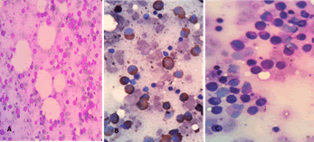
Investigations showed a Hemoglobin-6.6g/dl; TLC-3100/cumm; PLT-30,000/cumm. CT Brain showed subarachnoid haemorrhage of right frontal lobe and infarct in pericallosal region in left fronto-parietal lobe. Bone marrow examination revealed a hypercellular marrow with marked proliferation of Blasts and promyelocytes together constituting 48% (hyper granular variant) (Figure 1). On special staining the blasts were found to be positive for Sudan black while negative for Periodic acid Schiff stain (PAS). Final diagnosis of Acute Promyelocytic leukemia (Hypergranular variant) in a known case of carcinoma cervix was given.
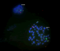
The diagnosis was again confirmed by Fluorescent in Situ Hybridization (FISH) which was found to be positive for t (15;17) PML-RARA Fusion (Figure 2). Patient was started on ATRA (All Trans Retinoic Acid) and Arsenic Trioxide. However, the patient’s clinical condition worsened with altered sensorium. Further treatment was not possible as the patient got discharged against medical advice.
Case-2:
A 14 years old female presented with pain in the right hip joint since 9 months. On clinical examination, the patient had a mass in the right iliac bone. On MRI, an infiltrative lesion involving right iliac bone was identified, which was later biopsied. A Diagnosis of Malignant Round cell tumour was made. Further, Immunohistochemistry was done with CD99 and vimentin positivity. Final Diagnosis of Ewing’s Sarcoma was made. She was started on chemotherapy for a period of 54 weeks. She also received radiotherapy between 15 to 24 weeks. Post chemotherapy, patient was on follow up and unremarkable.10 months after completion of treatment, patient presented with complaints of fever since 7 days.
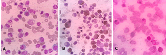
On investigating haemoglobin was found to be 4.1 gm/dl with a Total Leucocyte count of 3,00,000/cumm and Platelet count of 56,000/cumm. Bone marrow examination revealed a hypercellular marrow with Blast: 73, Myelocyte-11; Neutrophil-06, Lymphocyte-04, Monocyte-06. The blasts were Sudan black positive and negative for PAS (Figure 3). Final diagnosis of Acute Myelomonocytic Leukemia (AML-M4) in a known patient of Ewing’s Sarcoma was made.
Patient was started on hydroxyurea for acute leukemia. During treatment, patient developed Tumour Lysis Syndrome (TLS) with creatinine-8.5 mg/dl; Uric Acid-35mg/dl;Potassium-3.63 mmol/l; urine output-500 ml/24 hrs. To Alleviate the TLS patient underwent hemodialysis. However, the patient’s condition worsened with altered sensorium and right sided hemiplegia, due to intracranial bleeding and she expired.
Discussion
Secondary leukemia represents one of the most severe long term complications of cancer chemotherapy [2]. Various chemotherapeutic drugs such as alkylating agents, topoisomerases and platinum compounds induce leukemia. Cisplatinum-based concurrent chemoradiation is now the corner stone in the treatment of carcinoma cervix that has improved the survival [3]. Due to prolonged survival, treatment-related second malignancy is also increasingly being recognized. Platinum compounds produce intrastrand and interstrand DNA crosslinks [4]. The persistence of DNA adducts in the bone marrow might lead to secondary leukaemias. Radiation given with platinum compounds increases their carcinogenic potential [5]. This combination probably leads to the development of APML, in our first patient. In a similar case report by samanta et al, the second cancer developed after 63 months whereas the interval was just 17 months in our patient. Alkylating agents like Ifosfamide and Topoisomerase Inhibitors like Etoposide are used in treatment of Ewing’s Sarcoma. Alkylating agents have been implicated to cytogenetic alterations of chromosome 5 and/or 7 frequently progressing to AML-M1/M2 (French-American-British (FAB) classification). In contrast, topoisomerase inhibitors are associated with translocations of 11q23 resulting in AML-M4/5 [6]. In our second case of Ewing’s Sarcoma both these agents were used. Any of these would have been causative agent. However, the morphology was AML-M4.
Conclusion
We have documented two rare cases of Secondary Leukemias. These patients had a disease free survival following treatment of their primary malignancies. However, the Secondary Leukemias lead to poor prognosis.
References
- http://www.cancer.org/cancer/cancercauses/othercarcinogens/medicaltreatments/secondcancerscausedbycancertreatment/secondcancers-caused-by-cancer-treatment-intro
- KollmannsbergerC, BeyerJ, DrozJP, HarstrickA, HartmannJT, et (1998) Secondary leukemia following high cumulative doses of etoposide in patients treated for advanced germ cell tumors. J Clin Oncol 16: 3386–91.
[Crossref], [Google Scholar], [Indexed]
- Samanta DR, Senapati SN, Sharma PK, Mohanty A, Samantaray S, et al. (2009) Acute myelogenous leukemia following treatment of invasive cervix carcinoma: A case report and a review of the literature. J Cancer Res Ther 5: 302-304.
[Crossref], [Google Scholar]
- Greene MH(1992) Is cisplatin a human carcinogen. J Natl Cancer Inst 84: 306-12.
[Crossref], [Google Scholar]
- Vokes EE, Weichselbaum RR(1990) Concomitant chemoradiotherapy: Rationale and clinical experience in patients with solid tumors. J Clin Oncol 8: 911-34.
[Crossref], [Google Scholar], [Indexed]
- Pedersen-Bjergaard J, Philip P, Larsen SO, Jensen G, Byrsting K (1990) Chromosome aberrations and prognostic factors in therapy-related myelodysplasia and acute nonlymphocytic leukemia. Blood. 76: 1083–1091.
[Crossref], [Google Scholar], [Indexed]

Open Access Journals
- Aquaculture & Veterinary Science
- Chemistry & Chemical Sciences
- Clinical Sciences
- Engineering
- General Science
- Genetics & Molecular Biology
- Health Care & Nursing
- Immunology & Microbiology
- Materials Science
- Mathematics & Physics
- Medical Sciences
- Neurology & Psychiatry
- Oncology & Cancer Science
- Pharmaceutical Sciences


