Primary Hydatid Cyst of Infraspinatus Muscle
Mohammed Massine El-Hammoumi, Mohamed Montasser, Ayouba El-Hassane, Omar Slaoui, Fayçal El-Oueriachi and El-Hassane Kabiri
DOI10.21767/2471-8041.100036
Mohammed Massine El-Hammoumi1*, Mohamed Montasser2, Ayouba El-Hassane1, Omar Slaoui1, Fayçal El-Oueriachi1 and El-Hassane Kabiri1
1Faculty of Medicine and Pharmacy, Department of Thoracic Surgery, Mohamed V Military University Hospital, Morocco
2Faculty of Medicine and Pharmacy, Department of General Surgery, Avicenne Hospital, Ibn Sina University Hospital Centre, Mohamed V University, Morocco
- *Corresponding Author:
- Mohammed Massine El Hammoumi
Faculty of Medicine and Pharmacy
Department of Thoracic Surgery
Mohamed V Military University Hospital
Riad 10100 Rabat, Morocco
Tel: 00(212)666161916
Fax: 0033427869267
E-mail: hamoumimassine@hotmail.fr
Received date: July 12, 2016; Accepted date: December 23, 2016; Published date: December 26, 2016
Citation: El-Hammoumi MM, Montasser M, El-Hassane E. Primary Hydatid Cyst of Infraspinatus Muscle. Med Cas Rep. 2016, 2:4.. doi:10.21767/2471-8041.1000036
Abstract
Echinococcosis is an endemic zoonotic infection. The liver and the lung are the most affected organs, but muscle hydatidosis is very rare. We reported a case of a 39-yearold man, admitted for a cystic lesion of the right infraspinatus muscle, with radiological presumption of a hydatid cyst or a cystic tumor on computed tomography. Surgical excision of the cyst was performed. Preoperatively a diagnosis of an hydatid cyst was obvious. The pathologist confirmed the diagnosis.
Keywords
Echinococcosis; Soft tissue; Surgery
Introduction
Hydatid disease or echinococcosis is caused by the larval form of the cestode Echinococcus granulosus. It is an endemic zoonotic infection [1]. Hydatid cysts involved most frequently within the liver and lungs, but they are exceptionally identified in the soft tissues particularly skeletal muscles. To decline clinical confusion with other scapular masses we report a case of infraspinatus scapular hydatid cyst.
Learning Point for Clinicians
Soft tissue mass is not always a malignant tumor. Hydatid cyst should be included in the differential diagnosis in endemic areas especially that surgery can allow good control of the hydatid disease.
Case Presentation
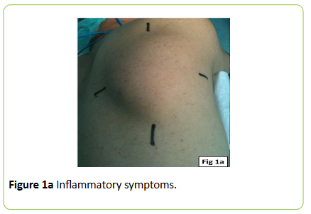
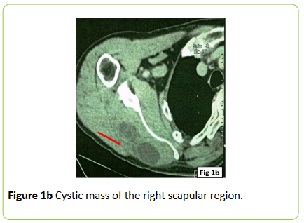
A previously healthy 39 years-old man, was admitted in our department for progressive growing painless mass discovered 22 months ago. On physical examination, a large right scapular cystic mass was noted. There was no local inflammatory symptoms (Figure 1a). A chest CT noted a well circumscribed cystic mass of the right scapular region (Figure 1b) containing partitions. A presumptive diagnosis of hydatid cyst of infraspinatus muscle was suspected.
Abdominal ultrasounds identified no other sites of the hydatid disease. Hydatid serological test was negative. Surgical resection of this mass was decided. Preoperatively, the cystic mass involved in the infraspinatus muscle and was surrounded by a thick fibrous capsule. Complete pericystectomy was safely performed. The macroscopic examination confirmed hydatid cysts with daughter vesicles.
The post-operative course was uneventful. The patient was discharged on the fourth postoperative day. Albendazole (400 mg twice a day) was prescribed for 3 months with). There was no recurrence after one year of following-up.
Discussion
Hydatid disease is a real problem of public health, especially in endemic areas such as North Africa, and the Middle East [2,3]. The most frequently affected organs are the liver (75% of cases) and lung (15%). Soft-tissue hydatid disease occurs in 2% or 3% of cases. Primary skeletal muscular hydatid cyst is rarely reported in the literature [3-6]. The pectoralis major, Sartorius, biceps brachii, supraspinatus and the neck were the most reported sites [1,4]. This involvement is possibly explained by the presence of lactic acid in the muscle. We report here another muscular involvement of hydatid disease.
Clinically diagnosis of intramuscular E. granulosus infection is difficult. It may resemble any soft-tissue tumor [1]. Serological tests and radiological examinations are very useful to the presumptive diagnosis [3]. MRI, ultrasound and CT can allow a definitive diagnosis of hydatid disease of muscle [1,2]. This preoperative evaluation is very important in order to avoid rupture, infection, anaphylaxis and recurrence of the disease [3].
Because hydatid cyst may mimic a solid soft-tissue mass, percutaneous biopsy or aspiration should not be performed because of the risk of dessimination or anaphylactic shock [2]. In our experience we first thought that was a subscapular elastofibroma but the cystic density in CT wasn’t presumptive of this diagnosis, besides some malignant tumor can contain cystic components.
Surgery should be undertaken safely to avoid iatrogenic rupture of the cyst [1,3]. The surgical field can be washed with hypertonic saline. Three months of albendazole is the preferred treatment protocol [1-3].
Conclusion
Muscular hydatid cyst is a rare entity, that can mimic a solid soft-tissue mass. Biopsy must be avoided and the preoperative diagnostic is based on radiological examinations. Surgical excision is the main modality of treatment.
References
- Tatari H, Baran O, Sanlidag T, Gore O, Ak D, et al. (2001) Primary intramuscular hydatidosis of supraspinatus muscle. Arch Orthop Trauma Surg121:93-94.
- Sreeramulu PN,KrishnaprasadS, Girish GSL (2010) Gluteal region musculoskeletal hydatid cyst: Case report and review of literature. Indian J Surg72:302-305.
- Abdel-Khalig RA, Othman Y (1986) Hydatid cyst of the pectoralis major muscle. ActaChirScand152: 469-471.
- Rask MR, Lattig GJ (1970) Primary intramuscular hydatidosis of the sartorius. J Bone Joint Surg Am 52:582-584.
- Duncan GJ, Tooke SMT (1990) Echinococcus infestation of the biceps brachii. ClinOrthop261:247-250.
- Eroglu A, Atabekoglu S, Kocaoglu H (1999) Primary hydatid cyst of the neck. Eur Arch Otorhinolaryngol256:202-204.

Open Access Journals
- Aquaculture & Veterinary Science
- Chemistry & Chemical Sciences
- Clinical Sciences
- Engineering
- General Science
- Genetics & Molecular Biology
- Health Care & Nursing
- Immunology & Microbiology
- Materials Science
- Mathematics & Physics
- Medical Sciences
- Neurology & Psychiatry
- Oncology & Cancer Science
- Pharmaceutical Sciences


