Neurofibromatosis Type-2: A Neurocutaneous Syndrome with Constellation of Multiple CNS Tumors
Rajendra Kumar Pandey, Ankur Garg and Durgvijay Singh
Rajendra Kumar Pandey*, Ankur Garg and Durgvijay Singh
Department of Neurology, Sawai Man Singh Medical College, Jaipur, India
- *Corresponding Author:
- Rajendra kumar pandey
Department of Neurology
Sawai Man Singh Medical College
Jaipur, India
Tel: 7727933909
E-mail: rp3556@gmail.com
Received date: January 05, 2016; Accepted date: April 05, 2016; Published date: April 10, 2016
Citation: Pandey RK, Garg A, Singh D. Neurofibromatosis Type-2: A Neurocutaneous Syndrome with Constellation of Multiple CNS Tumors. Med Case Rep. 2016, 2:2.
Abstract
Neurofibromatosis type-2 (NF-2) is a rare autosomal dominant hereditary disease caused by mutation of NF-2 gene on chromosome 22. Clinically it is characterized by presence of multiple CNS tumor types and is hence designated as central NF. Here we present a case of 20 year old male born of consainguous marriage who presented with multiple CNS tumors (vestibular schwannoma, meningioma, ependymoma and spinal neurofibroma) with juvenile posterior subcapsular cataract and café-au-lait spots. This case presents itself as a classic example of what has been described about NF-2.
Keywords
Vestibular schwannoma; Ependymoma; Spinal neurofibroma; Cutaneous and Skeletal deformity
Introduction
The neurofibromatosis consists of at least two distinct dominantly inherited disorders, neurofibromatosis type 1 (NF-1) and neurofibromatosis type 2 (NF-2). For many years both these conditions were considered as same. NF-2 was first described in 1822 by the Scottish surgeon, Wishart [1]. Heredity in NF-2 was first reported in 1920 by Feiling and Ward [2], In contrast to NF-1, there is paucity of cutaneous lesions, predominance of multiple CNS tumor types (especially vestibular schwannoma) and presence of posterior subcapsular cataract in NF-2.
Café-au-lait, also referred to as café-au-lait spots or caféau- lait macules are well-circumscribed, evenly pigmented macules and patches that range in size from 1 to 2 mm to greater than 20 cm in greatest diameter. While NF-1 is the most common syndrome associated with multiple café-au-lait, other syndromes associated with café-au-lait spots include McCune- Albright syndrome, Legius syndrome, Noonan syndrome and other neuro-cardio-facialcutaneous syndromes, ring chromosome syndromes, and constitutional mismatch repair deficiency syndrome. They are much less commonly found in NF-2 [3].
This patient is a 20 year old male who presented with all the classically described manifestations of NF-2 including multiple CNS tumor types, posterior subcapsular cataract, café-au-lait spots and skeletal deformity (scoliosis).
Case Report
A 20 year old male born of consaingous marriage (2nd degree) having normal birth and developmental history was presented with two year history of progressive hearing loss. The loss was initially on the left, followed by the right side and was associated with imbalance while walking and an occasional ringing sound in the ear. From the last one and a half year there was progressive painless diminution of vision in both eyes (left>right) to a current state of complete blindness. His attendants noticed a change in his voice from the last eight months which became slurred with nasal intonation. The patient also described difficulty in eating food as there was problem in manipulating food bolus in the mouth and an occasional chocking sensation from last four months.
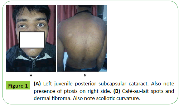
General physical examination revealed presence of café-au-lait spots (5 in number, over trunk), dermal fibroma over back and scoliosis (Figures 1A and 1B). The cranial nerve examination was remarkable showing decreased visual acuity to only perception of light in both eyes with fundus suggestive of secondary optic atrophy. Pupils were bilaterally mid-dilated and only sluggishly reacting to light. Cranial nerve examination revealed palsy of third, fourth, sixth and seventh cranial nerve on the right side and sixth cranial nerve on the left side. Eighth cranial nerve examination revealed bilateral sensor neural hearing loss with vestibular dysfunction. The examination also revealed palsy of ninth, tenth cranial nerve on right side and bilateral tweflth (left>right) cranial nerve with tongue fasciculation and atrophy. Motor examination showed pyramidal type of weakness in both the lower limbs with brisk DTR’s. There was only mild cerebellar dysfunction with grade 1 nystagmus.
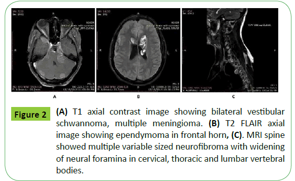
MRI brain with contrast revealed multiple variable sized extra axial well defined solid contrast enhancing mass in bilateral cerebello-pontine angle with widening of internal auditory canal (acoustic neuroma), falx cerebri, left cavernous sinus abutting left optic tract, both convexity of cerebellum, left parietal convexity, greater wing of sphenoid (suggestive of multiple meningioma) (Figure 2A). There was a mass lesion in the frontal horn of left lateral ventricle of heterogeneous intensity and heterogeneous enhancement with dilatation of lateral ventricle suggestive of ependymoma (Figure 2B). The MRI spine showed multiple variable sized neurofibroma with widening of neural foramina in cervical, thoracic and lumbar vertebral bodies (Figure 2C).
Figure 2: (A) T1 axial contrast image showing bilateral vestibular schwannoma, multiple meningioma. (B) T2 FLAIR axial image showing ependymoma in frontal horn, (C). MRI spine showed multiple variable sized neurofibroma with widening of neural foramina in cervical, thoracic and lumbar vertebral bodies.
Discussion
NF-2 is an autosomal dominant hereditary disease affecting 1 in 35000-50,000 people [4]. It is caused by mutation in NF-2 gene on chromosome 22 that codes for protein schwannomin or merlin [5]. Patients of NF-2 have paucity of cutaneous lesions and often have multiple types of CNS tumors (hence designated as central NF). Presence of bilateral acoustic neuroma, juvenile posterior subcapsular cataract, and other tumor types like meningioma, schwannoma, glioma or neurofibroma with positive family history is virtually diagnostic of NF-2 [6]. The term MISME syndrome (multiple inherited schwannomas, meningiomas, and ependymomas) applies to the disorder. Typical vestibular schwannoma arises from vestibular division of eighth nerve just within the internal auditory canal. As the tumor grows it occupies the angle between the cerebellum and pons. In this lateral position it is so situated that it can compress the seventh, fifth, and less often the ninth and tenth cranial nerves [7] Treatment of NF-2 requires multidisciplinary approach requiring a team of experienced neurosurgeon, otolaryngologist, audiologist, ophthalmologist, neuroradiologist, and geneticist. Small and stable asymptomatic acoustic schwannomas are followed with serial MRI scans on an annual basis. Sympatomatic tumors are treated with microsurgical resection or stereotactic radiosurgery. Surgical risk include postoperative CSF leak, facial and trigeminal neuropathy, and deafness. These risks are directly related to tumor size [8]. Bevacizumab therapy shrinks vestibular schwannomas and improves hearing in patients who have NF-2 [9]. Patients with damaged cochlear nerve can be rehabilitated successfully with a cochlear implant. Because detection of tumor at an early stage is effective in improving the clinical management of NF-2, presymptomatic genetic testing is an integral part of the management of NF-2 families.
Our patient, a 20 year old male presented with multiple CNS tumors (including bilateral acoustic neuroma, meningioma, ependymoma and multiple spinal neurofibromas) and posterior subcapsular cataract which is consistent with the diagnosis of NF-2. Although there was no family history and there were presence of multiple café-au-lait spots which are occasionally seen in NF-2. This case presents as a typical example of what has been described about NF-2.
References
- Wishart J (1822) Case of tumours in the skull, durameter, and brain. Edinburgh Med Surg J 18: 393-397.
- Feiling A, Ward E (1920) A familial form of acoustic tumour. BMJ 10: 496- 497.
- Shah N (2010) The diagnostic and clinical significance of café-au-lait macules. Pediatr Clin North Am 57: 1131-1153.
- Pollack J, John M (1997) Neurofibromatosis 1 and 2. Brain Pathol 7: 823- 836.
- Louis N, Ramesh V, Gusella F (1995) Neuropathology and molecular genetics of neurofibromatosis 2 and related tumours. Brain Pathol 5:163- 172.
- National Institutes of Health. Consensus Development Conference statement on neurofibromatosis. Neurofibromatosis Research Letter 1987 3: 3- 6.
- Stangerup (2012) Epidemology and natural history of vestibular schwannomas. Otolaryngol Clin North Am 45: 257- 268.
- Kaylie M, Gilbert E, Horgan A (2001) Acoustic neuroma surgery outcomes. Otol Neurotol 22: 686-689.
- Plotkin R, Stemmer-Rachamimov A, Barker G (2009) Hearing improvement after bevacizumab in patients with neurofibromatosis type 2. N Engl J Med 361: 358-367.

Open Access Journals
- Aquaculture & Veterinary Science
- Chemistry & Chemical Sciences
- Clinical Sciences
- Engineering
- General Science
- Genetics & Molecular Biology
- Health Care & Nursing
- Immunology & Microbiology
- Materials Science
- Mathematics & Physics
- Medical Sciences
- Neurology & Psychiatry
- Oncology & Cancer Science
- Pharmaceutical Sciences


