Vascular Malformation of Upper Lip ÃÆâÃâââ¬Ãâââ¬Å A Case Report
Chethana Sneha*, Imran Mohtesham and Priyal R
Department of Oral and Maxilofacial Pathology, Yenepoya Dental College, Yenepoya University, Mangalore, Karnataka, India
- *Corresponding Author:
- Chethana Sneha
Department of Oral and Maxilofacial Pathology, Yenepoya Dental College Yenepoya University
University Road, Deralakatte, Mangaluru, Karnataka 575018, India
Tel: 919964166164
E-mail: chethanasneha6164@gmail.com
Received Date: August 04, 2018; Accepted Date: August 28, 2018; Published Date: August 30, 2018
Citation: Sneha C, Mohtesham I, Priyal R (2018) Vascular Malformation of Upper Lip – A Case Report. Med Case Rep Vol.4 No.3: 83. DOI: 10.21767/2471-8041.100119
Abstract
Vascular anomalies (VM) account as one among the most difficult diagnostic and therapeutic enigmas that can be encountered in the practice of medicine. These lesions are the result of an embryonic abnormality of the vascular system. The main characteristic feature of vascular malformations is that they never show signs of involution. It is the most common neoplasm of the infancy. The clinical presentations are extremely protean and can range from an asymptomatic birthmark to life-threatening hemorrhage. Here we present a case of vascular malformation in the upper lip of four-year-old patient.
Keywords
Benign vascular lesions; Vascular malformation; Hemorrhage; Neoplasm
Introduction
Benign vascular lesions are abnormalities of blood vessel or endothelial cell proliferation. It was observed that benign oral vascular lesions represented 6.4% of all the diseases diagnosed by Oral Diagnosis Service [1,2]. Two most common types of vascular birthmarks are hemangiomas and vascular malformations which may appear to be very similar, but their course and treatment varies [3,4].
About 12% of newborns are thought to have a hemangioma, although most of them disappear during the first year of life instead vascular malformations are always present from birth, though they might not be apparent. Hemangiomas disappear with age but vascular malformations grow spontaneously with time. Hence, in general, most hemangiomas can be considered insignificant tumors that do not require any treatment except for aesthetic correction [4].
Based on the biological behaviors, VM can be subdivided into low flow and high flow lesions [5-7]. Oral vascular malformations are prevalent in the 6th and 7th decades of life. Histopathologically, it shows proliferation of endothelial cells with stasis of blood. Surgical excision followed by embolization is the treatment of choice for such lesions [8]. The commonest complication of such lesions is excessive bleeding during excision. Here we report a case of vascular malformation seen in upper lip of four and half year patient.
Case Report
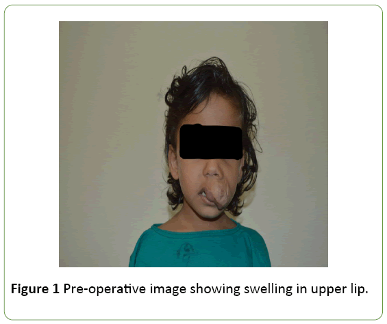
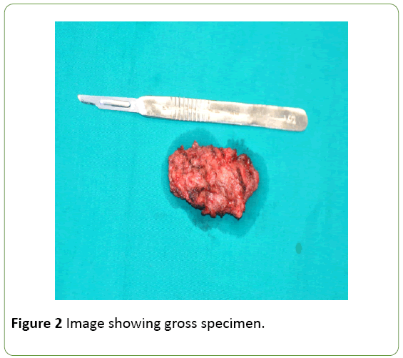
A four and half year-old patient had reported to OPD with a chief complaint of swelling in the left upper lip. The patient’s mother gives history of swelling seen after 6 months of birth which gradually increased in size with time. On clinical examination the swelling was large, sessile, measuring about 4 × 4 cm. The swelling was soft in consistency with irregular surface. The overlying mucosa was normal. Enlarged vein was visible, no pulsation present. There was no history of pain and paresthesia (Figure 1). On gross examination the specimen was measuring about 3 × 3 cm, reddish brown in color, soft in consistency (Figure 2).
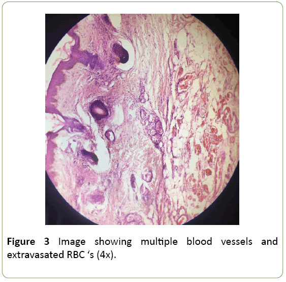
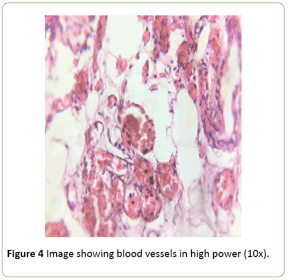
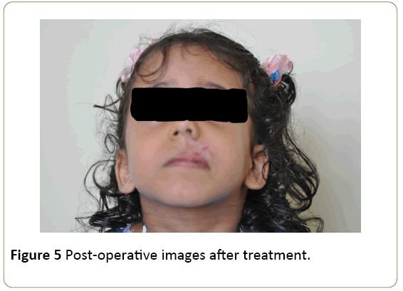
The histopathological feature showed connective tissue exhibiting multiple RBC’s filled with blood vessels of varying size and the nature ranging from small sized capillaries to medium sized arterioles which are interspersed with perinural arrangement of vascular channel suggestive of vascular malformation (Figures 3 and 4). The lesion was surgically excised (Figure 5) and patient was asked for follow up to check for recurrence.
Discussion
Benign vascular lesions are abnormalities of blood vessel or endothelial cell proliferation, it was observed that benign oral vascular lesions represented 6.4% of all the diseases diagnosed by Oral Diagnosis Service [2] Mulliken and Glowacki in 1982 proposed classification of vascular anomalies based on pathological features, i.e., endothelial cell turnover. According to this classification, vascular anomalies have been classified in to two categories - (a) Vasoproliferative neoplasm (b) Vascular malformations. Vascular malformation has less endothelial cell turnover (proliferates and undergo mitosis) compared to vasoprolifertive neoplasm. Instead VM are structural abnormalities of Venous, lymphatic, capillary and arterioles which grow according to the proportion of the child [9]. Enlargement of vascular lesions are due to changes in flow and pressure, dilatation of vascular channel, and collateral proliferation [8]. Active endothelial cells are the important feature in all series consistent with a progressing vascular lesion, but it is unclear whether the endothelial proliferation is a primary event or results from vascular expansion by means of hemodynamic mechanism [1]. Table 1 showing difference between haemangioma and vascular malformations.
| Hemangiomas | Vascular Malformation |
|---|---|
| A haemangioma may or may not be present at birth | Vascular malformation is always present at birth |
| They are true benign neoplasms of endothelial cells | Are localized defects of vascular morphgens that results in formation of abnormal torturous and enlarged vascular channel |
| Females are more commonly affected 3:1 (Mulliken and GLowacki) | Vascular malformations show no gender predilection |
| Hemangiomas are also known as port winestain, strawberry haemangioma, salmon patch. | Vascular malformations are also known as lymphangiomas, arteriovenous malformation, vascular gigantism. |
| Grows faster often faster than the child’s growth | Enlarges proportionately with growth of the child |
| Over time they become smaller (involute) and lighter in color. | They do not involute spontaneously and may become more apparent as child grows. |
| Mast cells known to play role in neangeogenisis, increases during proliferating phase. | No increase in mast cells. |
Table 1: Differences between haemangioma and vascular malformations.
VM are subdivided into (a) Slow-flow (b) High-flow malformations. Slow flow VM has prevalence of 1% in overall general population. Most common type in these subtypes is Venous, Lymphatic and Venolymphatic malformations. Venous malformation is formed due to dilatation of superficial and deep veins due to thin wall which lacks smooth muscle. Lymphatic subtypes of malformation are caused due to collection of lymph vessels filled with serous fluid. Venolymphatic malformations are rare [9].
High flow VM is anterivenous malformation and anterivenus fistula. They are characterized by formation of cluster of arterial and Venous channels without formation of solid mass. The clinical presentations exhibit extremely considerable variety and can range from an asymptomatic birthmark to lifethreatening haemorrhage. These lesions commonly occur in the head and neck region with a predilection for the oral cavity, respiratory and muscle groups. The overall incidence of VM is about 1 in 10000 people. They may continue to grow throughout the patient’s life. Many patients with vascular malformations may be misdiagnosed as hemangiomas [8].
Conclusion
Oral VM are more frequent in the upper lip, buccal mucosa and lower lip, doesn’t show any gender predilection [2]. Vascular malformations can cause significant morbidity and even mortality in both children and adults. Few syndromes such as blue rubber bleb nevus syndrome, cutaneo-mucosal venous malformation (VMCM), glomu-venous malformation (GVM) are associated with the vascular lesions [6]. Investigation includes MRI, CT and Doppler US. Benign oral vascular lesions may be treated by sclerotherapy, systemic corticosteroids, interferon α, laser, embolization, cryotherapy, and surgery. Management and treatment decisions depend on the patient’s age, and on the lesion’s site and size. The first line of treatment of VM includes embolization. Complete surgical excision or combination therapy is also suggested in children’s [9].
References
- Correa PH, Nunes LC, Johann AC, Aguiar MC, Gomez RS, et al. (2007) Prevalence of oral hemangioma, vascular malformation and varix in a Brazilian population. Braz Oral Res 21: 40-45.
- Redondo P (2007) Vascular malformations (I). Concept, classification, pathogenesis and clinical features. Actas Dermosifiliogr 98: 141-158.
- Foco F, Brkic A (2013) Vascular anomalies of the maxillofacial region: Diagnosis and management. Intechopen.
- Bhat VS, Arror R, Bhandary S, Shetty S (2013) Traumatic anteriovenous malformation of cheek: A case report and review of literature. Int J Ortholaryangol Clin 5: 173-177.
- Lowe LH, Marchant TC, Rivard DC, Scherbel AJ (2012) Vascular malformations: Classification and terminology the radiologist need to know. Semin Roentgenol 47: 106-117.
- Lakkasetty YK, Malik S, Shetty A, Nakhaei K (2014) Multiple vascular malformations in head and neck – A rare case report. Journal of Oral and Maxillofacial Pathology. J Oral Maxillofac Patho 18: 137-142.
- Barrett WA, Speight PM (2000) Superficial arteriovenous hemangioma of the oral cavity. Oral Surg Oral Med Oral Pathol Oral Radiol Endod 90: 731-738.
- Doifode D, Damle SG (2011) Vascular malformation of buccal mucosa: A case report. IJCD 2.
- Qiam F, Khan M, Din MQ (2011) Mucosal venous hemangiomas of the oral cavity – An analysis of 43 cases and literature review. JKCD 1: 2.

Open Access Journals
- Aquaculture & Veterinary Science
- Chemistry & Chemical Sciences
- Clinical Sciences
- Engineering
- General Science
- Genetics & Molecular Biology
- Health Care & Nursing
- Immunology & Microbiology
- Materials Science
- Mathematics & Physics
- Medical Sciences
- Neurology & Psychiatry
- Oncology & Cancer Science
- Pharmaceutical Sciences
