Tuberculosis of Calvaria - A Rare Entity
Swati Lalchandani, Raaghav Rai Verma, Rupak Kumar and Narender Singh Panghal
DOI10.21767/2471-8041.100065
1Department of Microbiology, Venkateshwara Hospital, Dwarka, New Delhi, India
2Department of Orthopaedics, Ram Manohar Lohiya Hospital & PGIMER, New Delhi, India
3Department of Orthopaedics, Employees State Insurance Hospital, ESI-PGIMSR Model Hospital, Basaidarapur, New Delhi, India
- *Corresponding Author:
- Rupak Kumar
Senior Resident, Department of Orthopaedics
Employees State Insurance Hospital, Baseaidarapur, New Delhi
Tel: 9599107680
E-mail: rupak.rims@gmail.com
Received Date: August 08, 2017; Accepted Date: September 22, 2017; Published Date: September 24, 2017
Citation: Lalchandani S, Verma RR, Kumar R, Panghal NS (2017) Tuberculosis of Calvaria - A Rare Entity. Med Case Rep, Vol.3 No.3:30. DOI: 10.21767/2471-8041.100065
Abstract
Tuberculosis is endemic in developing countries like India. However, calvarial tuberculosis is very rare. It was reported first time by Reid in 1942 and only a few cases have been reported till now. Most cases occur in first two decades secondary to haematogenous spread from a primary focus elsewhere in the body that may not always be evident. Calvarial tuberculosis in middle aged male with no evidence of tuberculosis elsewhere in body is very rare. Tuberculosis of the cranium is a rare entity, and can mimic tumors or multiple myeloma. A high index of suspicion and knowledge is required for an early diagnosis. We present a rare case of tuberculosis of calvaria in a middle-aged male who responded well to anti tubercular therapy.
Keywords
Tuberculosis; India; Calvarial; Cranium; Antitubercular therapy
Introduction
Tuberculosis continues to be among the greatest health problems in developing countries and has an enormous social and economic impact. During the past decade, the clinical pattern and presentation of tuberculosis has changed dramatically [1]. Since flat bones of skull contain little cancellous tissue, there is comparative rarity of the disease in skull. Tuberculosis of the flat bones of the vault of skull is rare and is considered secondary to an active or latent tuberculous lesion elsewhere in the body [2]. The first case of skull tuberculosis was described by Reid in 1842 [3]. In 1933, Strauss reviewed 220 cases of calvarial tuberculosis that had appeared in the literature and included three additional cases [4]. In 1942, Meng and Wu reported 40 cases in China of tuberculosis of the skull [5]. In 1981, Mohanty et al. reported 156 cases of chronic osteomyelitis of the skull in India, of which 22 cases were tuberculosis [6]. We are reporting a case of tuberculosis of the calvaria which is a rare entity.
Case Report
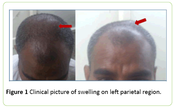
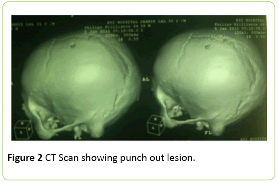
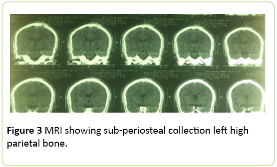
A 40-year-old male presented with swelling in the left parietal region for past one and half months. It was insidious in onset and gradually increasing in size. Patient had localized pain over the swelling. There was history of intermittent fever and loss of appetite for the same duration. On examination general condition of the patient was normal. Local examination showed a well demarcated bony hard swelling in the high left parietal region with dimensions of 5 × 5 cm (Figure 1). Local temperature was normal with no tenderness or erythema. Swelling was non- pulsatile and non expansile. Laboratory investigations revealed leucocytosis with predominantly lymphocytes and raised erythrocyte sedimentation rate of 45 mm in first hour the routine chest roentgenogram (PA view) and skull were normal. However, CT scan of skull revealed well circumscribed punched out lesion (size: 37 × 46 mm) in left high parietal region with bony fragment within with overlying soft tissue swelling with likely etiology of tuberculous osteomyelitis (Figure 2). MRI of the region revealed cortical defect measuring 9 mm with periosteal elevation and sub-periosteal collection on left high parietal bone (Figure 3). The findings were suggestive of osteomyelitis. FNAC was not attempted due the bony hard nature of the swelling.
An empirical diagnosis of tuberculous osteomyelitis of skull was made as invasive procedure was required to establish confirmed diagnosis of tuberculosis. Patient was started on 4 drug daily dose anti-tubercular therapy (Isoniazid 300 mg, Rifampicin 600 mg, Pyrazinamide 1500 mg, ethambutol 1000 mg) for two months. Patients symptoms responded to the therapy and the swelling disappeared after 2 months and his appetite improved. ATT was tolerated well by the patient. Post therapy MRI revealed resolution of the lesion.
Discussion
Before the advent of effective chemotherapy, calvarial tuberculosis was estimated to represent 0.2% to 1.3% of all cases of skeletal tuberculosis [4]. In his review, Strauss established an incidence of calvarial tuberculosis of 1%. In 1974, Nicholson reported only one case of calvarial tuberculosis in 176 cases of bone and joint tuberculosis [7]. Meng and Wu found both inner and outer tables to be equally affected, however Strauss reported affliction of inner table more likely [4].
Conclusion
Traditionally, surgical curettage was advised as a part of treatment [3], but now anti-tuberculosis therapy alone without surgery is treatment of choice. Here in this case, we treated a case of tuberculosis of calvaria in a middle-aged male who responded well to anti tubercular therapy.
References
- Glynn JR (1998) Resurgence of tuberculosis and the impact of HIV infection. Br Med Bull 54: 579-593.
- Gupta KB, Tandon S, Sen R, Kalra R (1998) Tuberculosis of the flat bone of the vault of skull - A case report. Ind J Tub 45: 231-232.
- Reid E (1998) MedizinichesCorrespondenzblattBayerischer Erlangen. 33: 1842.
- Strauss DC (1933) Tuberculosis of the flat bones of the vault of skull. SurgGynaecolObstet 57: 384-398.
- Meng CM, Wu YK (1942) Tuberculosis of the flat bones of the vault of the skull. J Bone Joint Surg 34: 341-353.
- Mohanty S, Rao CJ, Mukherjee KC (1981) Tuberculosis of the skull. IntSurg 66: 81-83.
- Nicholson RA (1974) Twenty years of bone and joint tuberculosis in Bradford: A comparison of the disease in the indigenous and Asian populations. J Bone Joint Surg 56B: 760-776F.

Open Access Journals
- Aquaculture & Veterinary Science
- Chemistry & Chemical Sciences
- Clinical Sciences
- Engineering
- General Science
- Genetics & Molecular Biology
- Health Care & Nursing
- Immunology & Microbiology
- Materials Science
- Mathematics & Physics
- Medical Sciences
- Neurology & Psychiatry
- Oncology & Cancer Science
- Pharmaceutical Sciences


