Personalized Imaging Approach in the Clinical Diagnosis of Prostate Cancer with Suspicion of Liver Metastases
Kalevi Kairemo, Kaarina Partanen, Anna ützow, Teppo Valle, Martti Ala-Opas and Timo Joensuu
Kalevi Kairemo1, Kaarina Partanen2, Anna ützow3, Teppo Valle3, Martti Ala-Opas4 and Timo Joensuu5*
|
| Corresponding Author: Timo Joensuu,Departments of Radiotherapy and Medical Oncology, Docrates Cancer Center, Helsinki, FI-00180, Finland.Tel: +358-10-773 2000 Fax: ++358-10-773 2099 E-mail: timo.joensuu@docrates.com |
| Received: October 24, 2015 Accepted: December 14, 2015 Published: December 28, 2015 |
| Related article at Pubmed, Scholar Google |
Visit for more related articles at Medical Case Reports
Abstract
The diagnostics of liver metastases in prostate cancer is challenging, while liver is seldom the primary site, and rarely the only primary metastatic organ.
Here we present two cases where liver metastases were suspected in prostate cancer patients. The differential diagnostics was successful by using 18F-choline PET/CT. The findings were confirmed by liver biopsy and consequent immunohistochemistry including PSA-staining. The first case verified liver as only metastatic site of prostate cancer and the second case appeared to be another primary cancer.
Keywords |
||||||||
| Prostate cancer; Liver metastases; Fluorocholine PET; Immunohistochemistry | ||||||||
Introduction |
||||||||
| Liver metastases are not rare in prostate cancer (PCa), in a large autopsy material of more than 1500 patients 25% cases had liver metastases [1] and in a large clinical material of more than 4000 patients liver metastases was found approximately in 4% of all cases [2]. In the large autopsy material bone (90%) and lung (46%) were more common sites for hematogenic metastatic spread [1]. But there are seldom conditions where the primary metastatic site is liver. In a French study of 345 patients with metastatic prostate cancer, six patients had liver metastases as the first site of metastatic disease, and for one of them, liver were the only metastatic site [3]. | ||||||||
| Choline for PET/CT is a routine diagnostic agent for prostate cancer which has European Medical Evaluation Agency (EMEA) approval in more than 20 European countries, and a recent meta-analysis in more than 3000 patients demonstrates its capability especially in nodal and metastatic PCa [4]. The sensitivity is dependent on Gleason score [5]. 18F-fluorocholine [chemical name known as [18F] fluoromethyl-dimethyl-2-hydroxyethyl-ammonium, (FCH)] shows particular promise as a PCa imaging agent [6-9]. | ||||||||
| At our institution we use FCH routinely for staging of prostate cancer [4] and radiation therapy (RT) dose planning of PCa [10,11]. | ||||||||
| We present two cases where liver metastases were suspected in prostate cancer patients. The differential diagnostics was successful by using 18F-choline PET/CT. The findings were confirmed by liver biopsy and consequent immunohistochemistry including PSA-staining. | ||||||||
Case Histories |
||||||||
| The first man was 59 year old having some urge without other urinary symptoms. His initial serum PSA was 9.8 μg/l/12% and clinically he had T3 disease in the left lobe of the prostate. In biopsies he had Grade 3, Gleason Score 7 (4+3), i.e. medium-high risk adenocarcinoma which covered 30% of the sample material. No metastases were seen in bone scintigraphy. Following radical prostatectomy and lymphadenectomy (9 obturatory lymph nodes from the left and 11 obturator lymph nodes from the right) the staging appeared to be T3bN0M0 and Gleason Score was confirmed to be 7. But due to the marginal positivity he received postoperative radiotherapy of 66 Gy in 2 Gy fractions to the prostate fossa. Following prostatectomy the AMS AdVance XP male sling operation was done due to the incontinence. New incontinence operation was planned but was cancelled due to the relapse of the prostate cancer. | ||||||||
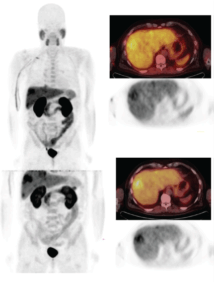
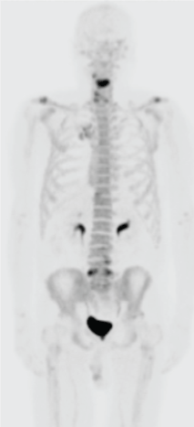
| 18 months after the prostatectomy the PSA was zero, but year later PSA increased up to 6.44 and patient became to Docrates Cancer Center where 18F-choline PET/CT and (sodium-18F-fluoride) Na18F-PET/CTs were performed. FCH-PET-study revealed four larger liver lesions, where SUVmax-values varied from 9.3 to 12.6 in the early images, and from 9.4 to 13.0 in the late images. Normally PCa spreads into the bone and/or local lymph nodes, but these regions did not demonstrate any pathologic findings (Figure 1). Also, the skeleton was free from metastatic findings, as Na18F-PET study demonstrates (Figure 2). | ||||||||
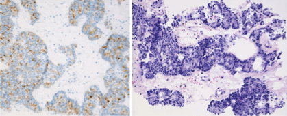
| Liver metastases were biopsied and the histological analysis revealed PSA positive prostate cancer (Figure 3). Docetaxel chemotherapy started. | ||||||||
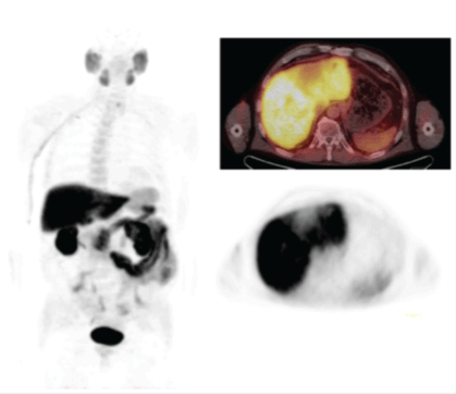
| The second man was 75 year old in good general condition who had regular clinical follow-up visits due to the mild unspecific kidney failure. Due to his age he wanted more complete urological check-up although he had no urological symptoms. Clear resistance in the apex of the prostate was observed by digital rectal examination and his initial PSA appeared to be elevated up to 10.4. In endorectal multiparametric MRI the size of the prostate was measured to be 36 g having some central hypertrophy. The tumor in the apex had ADC (apparent diffusion coefficient) of 0.7 x 10⁻³ mm2/s. In dynamic contrast enhanced images it was hypervascular and the tumor diameter was 1 cm. According to the radiologist’s analysis it looked more aggressive than Gleason Score 6 disease. Gleason Score 7 (3+4) diagnosis was also confirmed from biopsies. The cancerous transformation covered 10% of the biopsy sample. In choline- and Na18F-PET-CT we could not see any disseminated uptake of markers outside the prostate (Figure 4). However, in the liver there seemed to be unclear tumorous focus of 7 cm surrounding ligamentum falciforme. Based on abdominal CT the tumor in the liver was hypovascular arising suspicion more about cholangiocarcinoma than Hepatocellular Carcinoma (HCC). Tumor was biopsied and seemed to be PSA negative metastatic adenocarcinoma. The values of tumor markers were AFP 15 IU/l, CEA 3.6 μg/l and CA19- 9 4 IU/l. | ||||||||
| Patient received prophylactic single fraction irradiation of the mamillas and Degarelix + Bicalutamide started. | ||||||||
| Left lobe of the liver resected 3 months after primary prostate cancer diagnosis. The histopathological diagnosis of the liver tumor was confirmed to be cholangiocarcinoma and the resection was radical. | ||||||||
| T3N0M0 prostate cancer radiotherapy with VMAT RapidArc technique was given 5-7 months following initial diagnosis as image guided radiotherapy giving 45/1.8 Gy to the pelvic lymph nodes, 50 Gy to the seminal vesicles and 80 Gy in 2 Gy fractions to the prostate. Hormone therapy was not continued after radiotherapy. | ||||||||
| Written informed consent was obtained from both of the patients for publication of this case report and accompanying images. Copies of the written consents are available for review by the Editor-in-Chief of this journal. | ||||||||
Discussion |
||||||||
| Liver metastases are uncommon although not very rare, but appear to be a rather late event in the course of the disease [2]. They are frequently associated with neuroendocrine characteristics [2,3]. Hematogeneous metastases were present in 35% of 1,589 patients with prostate cancer, with most frequent involvement being bone (90%), lung (46%), liver (25%), pleura (21%), and adrenals (13%) [1]. | ||||||||
| In the literature one example with prostate liver metastases, C-11- choline [12] was not able to demonstrate metastatic liver disease, in contrast to our findings. C-11 has a short half-life, 20 min, which is a drawback in the diagnosis. In normal metabolism choline in the liver gives some background, but it decreases significantly already e.g. at 60 min. The lesions are clear in Figure 1. | ||||||||
| It is known from the literature that hepatocellular carcinoma and similar conditions are hypometabolic, i.e. cause a defect in the FCH-PET-studies [13]. Also, quantitative differential diagnosis of hepatocellular adenoma and focal nodular hyperplasia is possible using 18F-fluorocholine PET/CT based on hypometabolism degree [14]. | ||||||||
| FDG may demonstrate liver metastases [15], but FDG is not a good tracer for PCa [4]. There are other tracers [11,16], which may play pivotal role in the dsifferential diagnosis of PCa by molecular imaging -one of them is 68Ga-PSMA. In the literature a case is presented where 68Ga-PSMA was able to detect liver metastases [17]. We also know that FCH is helpful in the differential diagnosis of liver metastases in PCa, especially if late imaging is applied to improve the signal in liver (Figure 1). FCH is known to specific for prostate cancer: The sensitivity of FCH was 46-49%, whereas specificity was 92-95% in two large reviews of more than 2000 patients [4,18]. | ||||||||
| At this moment FCH-imaging is considered as a standard method in the staging and detection of biochemical relapse in PCa. Thousands of patients have been studied (4,5). Very little is known about detection liver metastases. Therefore, we recommend that late imaging covers liver region and it should be performed at least 60 minutes after injection. With 11C liver metastasis detection might be problematic, because of its short half-life of 20 minutes very late imaging is not possible. | ||||||||
Conflicts of Interest |
||||||||
| The authors declare no conflicts of interest. | ||||||||
Figures at a glance |
||||||||
|
||||||||
References |
||||||||
|
Select your language of interest to view the total content in your interested language

Open Access Journals
- Aquaculture & Veterinary Science
- Chemistry & Chemical Sciences
- Clinical Sciences
- Engineering
- General Science
- Genetics & Molecular Biology
- Health Care & Nursing
- Immunology & Microbiology
- Materials Science
- Mathematics & Physics
- Medical Sciences
- Neurology & Psychiatry
- Oncology & Cancer Science
- Pharmaceutical Sciences


