Massive Posterior Mediastinal Goiter Managed by ranscervical and Right Uniportal (Single Incision) Approach
Nardini Marco, Valeriya Okatyeva, Matteo A Cannizzaro and Migliore Marcello
DOI10.21767/2471-8041.S1-010
Nardini Marco, Valeriya Okatyeva, Matteo A Cannizzaro and Migliore Marcello*
Department of Thoracic and Endocrine surgery, Policlinico University Hospital, University of Catania, Via Santa Sofia,78, Catania 95100, Italy
- *Corresponding Author:
- Migliore Marcello
Department of Thoracic and Endocrine surgery
Policlinico University Hospital, University of Catania
Via Santa Sofia,78, Catania 95100, Italy
Tel: +39 095 743 1111
E-mail: mmiglior@unict.it
Received Date: January 18, 2018; Accepted Date: January 27, 2018; Published Date: January 29, 2018
Citation: Marco N, Okatyeva V, Cannizaro MA, Marcello M (2018) Massive Posterior Mediastinal Goiter Managed by Transcervical and Right Uniportal (Single Incision) Approach. Med Case Rep Vol.4 No. S1:010. DOI: 10.21767/2471-8041.S1-010
Abstract
The present case is of a 55-year-old female who underwent right hemi-thyroidectomy for a benign multinodular goiter 19 years before. Computed tomography showed the presence of a large posterior mediastinal mass which extended from the neck to the level of the right main bronchus adherent to the boyden branch of the right pulmonary artery. The patient underwent thyroidectomy through cervicotomy and right uniportal anterolateral thoracotomy. The mass was dissected free and then successfully removed. The patient was discharged 12 days post-operatively. There were no signs of recurrence at 6-year follow-up.
Keywords
Mediastinum; Surgery; VATS; Uniportal; Single Incision; Goiter
Case Presentation
A 55-year-old female presented with productive cough without dyspnea. Medical history included a right hemithyroidectomy for a benign multinodular goiter 19 years before. On physical examination there was a diffuse thyroid enlargement suggesting a recurrent goiter.
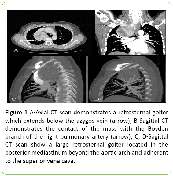
Laboratory tests and the thyroid function tests were within normal limits. Computed tomography (CT) showed the presence of a posterior mediastinal mass measuring 100 × 82 × 56 mm which extended from the neck retrotracheally beyond the aortic arch to the level of the right main bronchus adherent to the boyden branch of the right pulmonary artery (Figure 1). The trachea together with esophagus was deviated to the left. The arch of the azygos vein was restricted and displaced downwards.
Figure 1: A-Axial CT scan demonstrates a retrosternal goiter which extends below the azygos vein (arrow); B-Sagittal CT demonstrates the contact of the mass with the Boyden branch of the right pulmonary artery (arrow); C, D-Sagittal CT scan show a large retrosternal goiter located in the posterior mediastinum beyond the aortic arch and adherent to the superior vena cava.
The patient underwent thyroidectomy, but it was impossible to remove the mass through the transcervical incision. A 5cm right uniportal anterolateral approach was performed. The large thyroid mass was identified, and its lower edge was found below the level of the arch of the azygos vein.
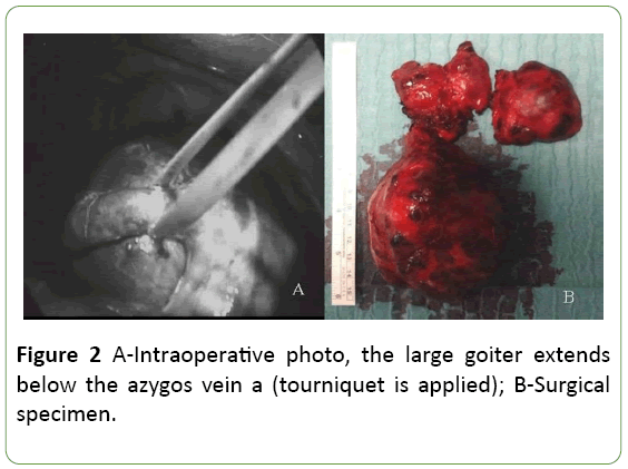
The mediastinal pleura were opened below the azygos vein from where the dissection was initiated (Figure 2). We had some difficulties with isolation of the mediastinal portion of the thyroid gland because there were many adhesions to the azygos vein, main pulmonary artery, the trachea and superior vena cava. During the preparation we found some additional vessels which were clipped and cut. The mass was dissected free and then successfully removed. Moreover, due to difficulty weaning the patient from the ventilator, he was transferred to the intensive care unit. After 3 days a tracheostomy was performed because difficulty in weaning and dense secretions. Patient was discharged 12 days postoperatively. The patient did not experience postoperative complications such as transient hypocalcemia, recurrent laryngeal nerve injury, bleeding, wound infection, or tracheomalacia. Postoperative pathology examination revealed a benign multinodular goiter. There were no signs of recurrence at 6-year follow-up.
The definition of mediastinal goiter is still not standardized. Most commonly, the goiter is defined mediastinal when it extents below the thoracic inlet or when more than 50% of the thyroid tissue is located inferior to this level [1]. Mediastinal goiter is most frequently situated in the anterior mediastinum [2]. Only 10–15% of intrathoracic goiters are located in the posterior mediastinum and their incidence is higher in patients with a history of previous thyroid surgery [3]. The mediastinal goiters are most often found in women than in men (female/ male ratio is about 3:1) [2]. Huins et al. [4] have proposed a classification of intrathoracic goiters based on the anatomical location and the type of operative approach. Mediastinal goiters more often than cervical goiters cause obstructive symptoms such ascough, dyspnea, stridor and dysphagia. Patients with mediastinal goiter which extend beyond the aortic arch o reach the level of the carina tracheae have not only an increased risk of intraoperative o postoperative complications such as recurrent laryngeal nerve damage, bleeding, transient or persistent hypoparathyroidism, pneumothorax, pneumonia, tracheomalacia but also increased mortality [2,5].
Discussion
Surgical management of posterior mediastinal goiters and mass can pose considerable problems [6,7]. Surgery is indicated in the presence of compressive symptoms o when the malignancy is suspected [2,8]. Some authors consider that mediastinal goiters should be treated surgically independent of clinical symptoms because of high incidence of malignancy and neoplastic transformation [3,9].
Malignancy has been reported in 1.4% to 21% of patients with mediastinal goiters. Although most of the mediastinal goiters can be managed by a transcervical approach [2,3], 2% to 29% of patients need an additional incision such as manubriotomy, sternotomy, thoracotomy. Most often the indications for additional incision include the large size, involvement of the posterior mediastinum, malignancy, extension below the aortic arch, airway obstruction and intraoperative complications [2,10]. In a study conducted in 2426 patients who underwent thyroidectomy, the transcervical approach was used in 84% of cases, manubriotomy in 3,1%, full sternotomy in 6,6%, and full thoracotomy in 4% [4]. Furthermore, the attempts to remove some posterior mediastinal goiters only by transcervical approach can lead to serious hemorrhage and injury to the adjunct structures especially in patients with a history of previous thyroidectomy [6].
Although videomediastinoscopy and video assisted thoracoscopy (VATS) approach can be used to remove mediastinal goiter [10,11] in the case of massive lesions, especially if the thyroid mass is located in the posterior mediastinum or there are tight adhesions to the adjacent structures, this approach becomes technically demanding.
Conclusion
In conclusion, transcervical and uniportal (single incision) video-assisted thoracic approach [12] can be used for large posterior mediastinal goiter delivery. This approach offers several additional advantage such us better visualization of the operative field, control of possible intraoperative hemorrhage, and easily extraction of the enlarged thyroid mass.
References
- Ríos A, Rodríguez JM, Balsalobre MD, Tebar FJ, Parrilla P (2010) The value of various definitions of intrathoracic goiter for predicting intra-operative and postoperative complications. Surgery 147: 233-238.
- Machado NO, Grant CS, Sharma AK, Sabti HA, Kolidyan SV (2011) Large posterior mediastinal retrosternal goiter managed by a transcervical and lateral thoracotomy approach. Gen Thorac Cardiovasc Surg 59: 507-511.
- Hajhosseini B, Montazeri V, Hajhosseini L, Nezami N, Beygui RE (2012) Mediastinal goiter: a comprehensive study of 60 consecutive cases with special emphasis on identifying predictors of malignancy and sternotomy. Am J Surg 203: 442-447.
- Huins CT, Georgalas C, Mehrzad H, Tolley NS (2008) A new classification system for retrosternal goitre based on a systematic review of its complications and management. Int J Surg 6: 71-76.
- Sancho JJ, Kraimps JL, Sanchez-Blanco JM, Larrad A, Rodríguez JM, et al. (2006) Increased mortality and morbidity associated with thyroidectomy for intrathoracic goiters reaching the carina tracheae. Arch Surg 141: 82-85.
- Landreneau RJ, Nawarawong W, Boley TM, Johnson JA, Curtis JJ (1991) Intrathoracic goiter: Approaching the posterior mediastinal mass. Ann Thorac Surg 52: 134-135.
- Nardini M, Jayakumar S, Migliore M, Dunning J (2017) Videoassisted thoracic surgery mediastinal germ cell metastasis resection. Interact Cardiovasc Thorac Surg 25: 160-161.
- Shahian DM, Rossi RL (1988) Posterior mediastinal goiter. Chest 94: 599-602.
- Mack E (1995) Management of patients with substernal goiters. Surg Clin North Am 75: 377-394.
- Agha A, Glockzin G, Ghali N, Iesalnieks I, Schlitt HJ (2008) Surgical treatment of substernal goiter: An analysis of 59 patients. Surg Today 38: 505-511.
- Migliore M, Costanzo M, Cannizzaro MA (2010) Cervicomediastinal goiter: Is telescopic exploration of the mediastinum (video mediastinoscopy) useful?. Interact Cardiovasc Thorac Surg 10: 439.
- Al-Mufarrej F, Margolis M, Tempesta B, Strother E, Gharagozloo F (2010) Novel thoracoscopic approach to difficult posterior mediastinal tumors. Gen Thorac Cardiovasc Surg 58: 636-639.

Open Access Journals
- Aquaculture & Veterinary Science
- Chemistry & Chemical Sciences
- Clinical Sciences
- Engineering
- General Science
- Genetics & Molecular Biology
- Health Care & Nursing
- Immunology & Microbiology
- Materials Science
- Mathematics & Physics
- Medical Sciences
- Neurology & Psychiatry
- Oncology & Cancer Science
- Pharmaceutical Sciences


