Extensor Tendon Rupture in Non-Traumatic Osteoarthritis of Distal Radioulnar Joint: Vaughan-Jackson Syndrome ÃÆâÃâââ¬Ãâââ¬Å A Rare Case Report from Indian Subcontinent
Rajesh Lalchandani
DOI10.21767/2471-8041.100071
Department of Orthopedics, ESI-PGIMSR Model Hospital, Basaidarapur, New Delhi, India
- *Corresponding Author:
- Rajesh Lalchandani
Director, Department of Orthopedics, ESI-PGIMSR Model Hospital, Basaidarapur, New Delhi, India
Tel: +011 2510 0664
E-mail: rupak.rims@gmail.com
Received Date: July 31, 2017, Accepted Date: August 20, 2017, Published Date: August 22, 2017
Citation: Lalchandani R (2017) Extensor Tendon Rupture In Non-Traumatic Osteoarthritis of Distal Radioulnar Joint:Vaughan-Jackson Syndrome– A Rare Case Report From Indian Subcontinent. Med Case Rep, Vol.3 No.4:36. doi: 10.21767/2471-8041.100071
Abstract
Osteoarthritis of distal radioulnar joint with rupture of extensor tendon of hand is a rare problem seen in elderly. It was first reported by Vaughen and Jackson in 1948 and the syndrome is named after them. The rupture results from attrition of dorsally dislocated ulnar head during pronation and supination movements. This dislocation of ulnar head is due to dehiscence of dorsal capsule of the distal radioulnar joint. “Scallop sign”1 is a radiological sign which are seen in this condition and signifies impending rupture of extensor tendon. Tendon transfer with excision of ulnar head is a viable option. We present a case of attritional rupture of extensor tendon of 4th and 5th fingers in a patient with non-traumatic osteoarthritis of distal radioulnar joint (DRUJ) in an elderly female. To the best of our knowledge no case of attritional rupture of extensor tendon of hand with osteoarthritis of distal radioulnar joint has been reported from Indian subcontinent.
Keywords
Distal Radioulnar joint; Rupture; Osteoarthritis; Extensor tendon; Scallop sign
Introduction
Extensor tendon rupture in association with osteoarthritis of the distal radioulnar joint (DRUJ) is a very rare entity and sporadically reported [1-4] although spontaneous extensor tendon rupture is known to occur in patients with rheumatoid arthritis [1,2]. It is generally due to dorsal dislocation of ulnar head caused by rupture of dorsal capsule of DRUJ [3-5] resulting in attrition of extensor tendon on pronation and supination movement. We present a case of extensor tendon rupture in a patient with osteoarthritis of distal radioulnar joint with the intention to discuss the mechanism and risk factors of this rare entity.
Case Presentation
A 60-year-old right hand dominant woman presented in our OPD with complaints of inability to extend her right little and ring finger for three months. Initially little finger was involved and a month later the ring finger also got involved. This was not associated with any trauma or pain. Patient worked in a button making factory for thirty years.
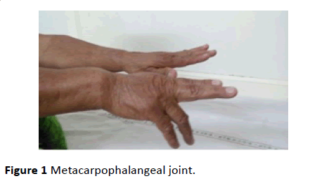
Thorough examination revealed a swelling over the dorsal aspect of wrist on the ulnar side. Patient was unable to actively extend her right little and ring fingers at the metacarpophalangeal joint (Figure 1).
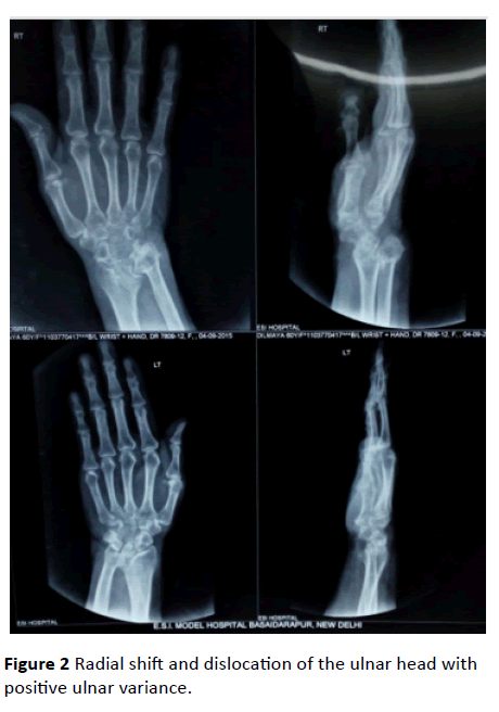
DRUJ was found to be unstable on load and shift tests. Blood cell count, erythrocyte sedimentation rate, and C-reactive protein were within normal parameters. Rheumatoid factor as well as Anti-CCP was negative. A study of the plain radiographs revealed osteoarthritic changes at the DRUJ with deepening and widening of the sigmoid notch (Scallop sign). Radial shift and dislocation of the ulnar head with positive ulnar variance was present (Figure 2).
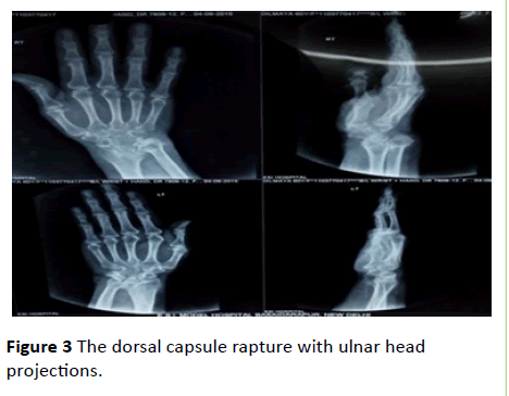
Intraoperatively, extensor digiti minimi tendon, along with the 4th and 5th extensor digitorum communis tendons were found to be ruptured with their distal ends lying engulfed in a matted mass. The proximal ends were retracted and were lying at the level of the wrist. The dorsal capsule was ruptured with ulnar head projecting from it (Figure 3). ‘Pannus’ was not seen which a characteristic finding of rheumatoid arthritis.
As the margins of the ruptured tendons were frayed and retracted till the wrist joint, end to end repair was not possible. The only feasible plan was to transfer the extensor indicis proprius tendon to the extensor digiti minimi tendon and to suture the distal stump of fourth and fifth EDC tendon with intact EDC tendon. The dislocated arthritic ulnar head was resected. The diseased synovium was sent for histopathological examination which was negative for rheumatoid arthritis.
At six months after surgery, patient was able to extend her ring and little finger without any difficulty (Figure 4). Minimal occasional pain is present around the wrist area.
Discussion
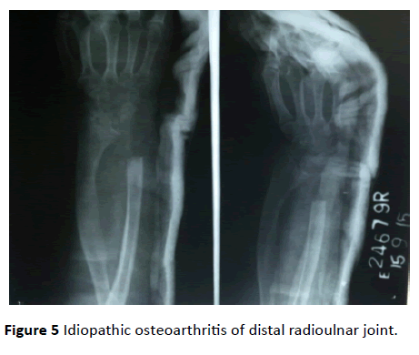
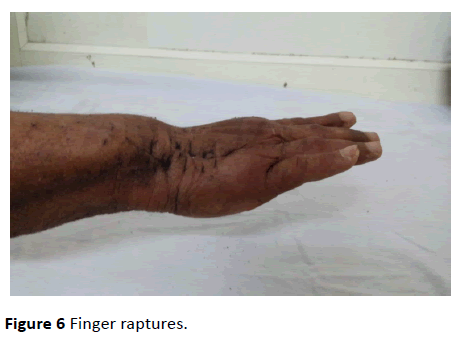
Idiopathic osteoarthritis of distal radioulnar joint is a rarity as revealed by review of literature (Figure 5). It was in 1948, when Vaughan-Jackson [2] for the first time reported two cases of attritional extension tendon rupture due to osteoarthritic distal radioulnar joint. Initially little finger rupture takes place followed by ring finger (Figure 6). This attrition is a result of friction between tendon and dorsally dislocated ulnar head and generally take place in the order of little finger first followed by ring finger. Radiologically, the reported finding is widening of the sigmoid notch (Scallop sign), shift of radial head radially, dorsal subluxation of ulnar head. ‘Scallop sign’ was first described by Frieberg and Weinstein [1] and is characterized by sclerotic border with deepening and widening of sigmoid notch. Its importance lies in the fact that it is prognostic indicator of impending extensor tendon rupture as reported by Yamazaki and colleagues [5].
Conclusion
Ohshio [3] and colleagues have reported five cases of attritional rupture of the extensor tendon due to osteoarthritis of the DRUJ. Carr and Burge [4] described the importance of dislocation of ulnar head as a result of perforation of dorsal capsule. According to them, this is the cause of attrition resulting in rupture in all cases. Our findings are consistent with few case reports which have been reported in literature.
References
- Freiberg RA, Weinstein A (1972) The scallop sign and spontaneous rupture of finger extensor tendons in rheumatoid arthritis. Clin Orthop Relat Res 7: 128-130.
- Vaughan-Jackson OJ (1948) Rupture of extensor tendons by attrition at the inferior radio-ulnar joint; Report of two cases. J Bone Joint Surg 7: 528-530.
- Ohshio I, Ogino T, Minami A, Kato H, Miyake A (1991) Extensor tendon rupture due to osteoarthritis of the distal radio-ulnar joint. J Hand Surg 7: 450-453.
- Carr AJ, Burge PD (1992) Rupture of extensor tendons due to osteoarthritis of the distal radio-ulnar joint. J Hand Surg 7: 694-696.
- Yamazaki H, Uchiyama S, Hata Y, Murakami N, Kato H (2008) Extensor tendon rupture associated with osteoarthritis of the distal radioulnar joint. J Hand Surg 7: 469-474.

Open Access Journals
- Aquaculture & Veterinary Science
- Chemistry & Chemical Sciences
- Clinical Sciences
- Engineering
- General Science
- Genetics & Molecular Biology
- Health Care & Nursing
- Immunology & Microbiology
- Materials Science
- Mathematics & Physics
- Medical Sciences
- Neurology & Psychiatry
- Oncology & Cancer Science
- Pharmaceutical Sciences
