Bilateral Pathological Fracture of Proximal Femur Revealing a Rare Lung Carcinoma: What the Family Physician Should Know?
Naoufal Elghoul, Azzelarab Bennis, Nasser Ibo, Abdeloihab Jaafar and Mohammed Tbouda
Department of Orthopedic Surgery and Traumatology, Military Hospital Mohamed V (HMIMV), Rabat, Morocco
- Corresponding Author:
- Elghoul Naoufal
Department of Orthopedic Surgery and Traumatology, Military Hospital Mohamed V (HMIMV), BP 10100 Rabat, Morocco
Tel: +2125377-14419
E-mail: naoufal.elghoul@gmail.com
Rec date: January 14, 2019; Acc date: January 31, 2019; Pub date: February 02, 2019
Citation: Naoufal E, Azzelarab B, Nasser I, Mohammed T, Abdeloihab J (2019) Bilateral Pathological Fracture of Proximal Femur Revealing A Rare Lung Carcinoma: What the Family Physician Should Know? Med Case Rep Vol. 5 No.4: 108. doi: 10.21767/2471-8041.100130.
Abstract
Lung cancer is a severe disease often diagnosed a late stage when surgical resection is no longer possible. Adenosquamous carcinoma is a rare subtype of lung carcinoma. It behaves more aggressively than other histologic subtypes of lung cancer with a poor long-term prognosis. We present here the case of a 62-years-old elderly male patient, smoker with chronic obstructive pulmonary disease, consulted initially a family physician for dyspnea, cough and a spontaneous pain of both hips then he was diagnosed as simple exacerbation of his pulmonary disease. Three months later, patient presented a bilateral pathological fracture of proximal femur prompting the patient to undergo surgery. He benefited from bilateral long Gamma intramedullary nail along with biopsy and cementation on the right proximal femur. Based on clinical features, chest x-ray finding, CT scan, histopathological finding and immune-histochemical analysis, a diagnosis of metastatic adenocarcinoma of femur, primary from the lung was made and was transferred to the oncology service for inpatient palliative radiation and chemotherapy. But, at 3 weeks from surgery, patient presented an acute decompensation of his chronic obstructive bronchopneumopathy causing his death.
Keywords
Pathological fracture; Lung carcinoma; Adenosquamous; Proximal femur
Introduction
Lung cancer is a severe disease often diagnosed a late stage when surgical resection is no longer possible because of local advancement or distant metastasis [1]. The spine and ribs are often the earliest sites of bone metastases, whereas the skull, femur, humerus, and scapula are involved later [2]. Adeno squamous carcinoma is a rare subtype of lung carcinoma, which accounts for 0.4% to 4% of all lung carcinomas [3]. A pathological fracture as the initial presentation of primary adeno squamous carcinoma is very rare [4] and only few cases are reported in literature [5].
To the best of our knowledge, the case we present here is among the rare case of unusual presentation of adeno squamous carcinoma in lung which illustrate the gravity if family physician missed the diagnosis.
Case Presentation
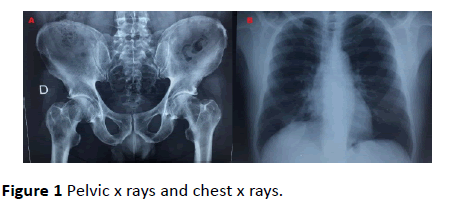
In late March 2018, A 62-years old elderly male patient, smoker (60 packs a year) with chronic obstructive pulmonary disease consulted a family physician for dyspnea, cough and a spontaneously pain of both hips for 1 month; without history of trauma or prior fracture. The right hip was more painful. A chest and pelvic x ray-s were realized. The physician has concluded to simple exacerbation of chronic obstructive pulmonary disease with no visible fracture on x ray-s (Figure 1). A paracetamol with salbutamol associated to antibiotic were prescribed and rest advised.
Three months later, the pain increased steadily becoming very intense contemporary to an audible crunch in the right hip causing total functional impotence of the right lower limb. Then the patient was transferred to the emergency department.
On arrival, the patient was in mild distress because of pain. Temperature 37.7°C, his blood pressure was 107/68 mmHg, pulse rate 96 beats/min, respiratory rate 18 breaths/min, SaO2 90% on room air without signs of respiratory distress and was in poor general health. Physical examination was significant for diminished lung sounds over the right chest. The right lower limb was on external rotation with palpation and mobilization of the right hip were painful along with painful left hip its mobilization was possible but limited.
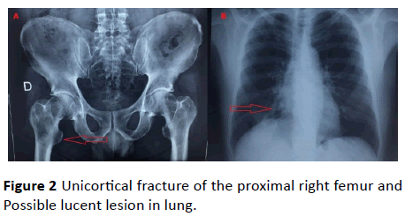
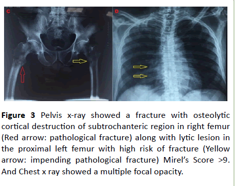
(At this follow up of visit, the patient reported very little subsequent benefit from prescribed treatment along with weight loss and persistence of hips pain with no aggravation of cough and dyspnea. His only relief came from limitation of weight bearing on both hips using the crutches. The review of the initial chest and pelvic x ray-s did show the presence of fracture in the proximal right femur and did suggest a lucent lesion in lung (Figure 2). The recent pelvic x ray (Figure 3) showed a fracture with osteolytic cortical destruction of subtrochanteric region in right femur (pathological fracture) along with lytic lesion in proximal left femur with high risk of fracture (impending pathological fracture) Mirel’s Score was more than 9 prompting the patient to undergo surgery (Table 1).
Figure 3 Pelvis x-ray showed a fracture with osteolytic cortical destruction of subtrochanteric region in right femur (Red arrow: pathological fracture) along with lytic lesion in the proximal left femur with high risk of fracture (Yellow arrow: impending pathological fracture) Mirel’s Score >9. And Chest x ray showed a multiple focal opacity.
| Score | Site of lesion | Size of lesion | Nature of lesion | Pain |
|---|---|---|---|---|
| 1 | Upper limb | < 1/3 of cortex | Blastic | Mild |
| 2 | Lower limb | 1/-2/3 of cortex | Mixed | Moderate |
| 3 | Trochanteric region | >2/3 of cortex | Lytic | Functional |
Table 1 Mirel’s scoring system; ≥ 9 prophylactic fixation.
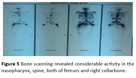
Chest X ray showed a multiple focal opacities (Figure 3) that shaped the need of a whole-body dynamic scan that revealed a mass in lower right lung measuring 70 × 40 mm with local extension and secondary localizations: liver, spine, and nasopharynx. Bone scanning revealed considerable activity in the nasopharynx, spine, both of femur and right collarbone.
a mass in lower right lung measuring 70 × 40 mm with local extension and secondary localizations: liver, spine, and nasopharynx. Bone scanning revealed considerable activity in the nasopharynx, spine, both of femur and right collarbone.
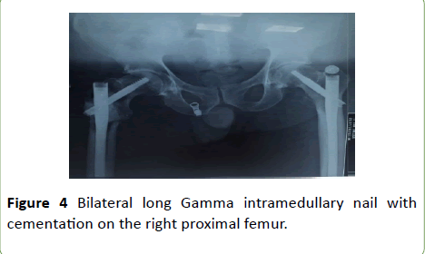
5 days later, after anesthetic preparation, he was admitted to the operating room. Thus, under general anesthesia, using a special orthopedic table, he benefited from long Gamma intramedullary nail in both proximal femurs along with biopsy and cementation on the right femur: the right side and left side were operated respectively (Figure 4). A clinical assessment of the patient 3 days post-operative showed that he presented no signs of respiratory or hemodynamic distress, no pain (under simple paracetamol) and possible weight bearing on both hips using 2 crutches.
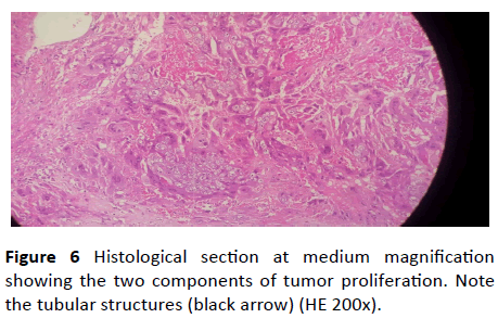
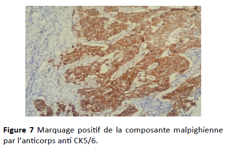
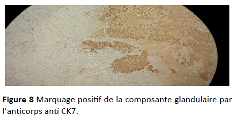
Histological study found a dual-component tumor proliferation: A glandular component made of tubular structures of variable size surrounded by cells with moderate atypia, and a squamous component made of guts within a desmoplastic stroma Figure 5. An immunohistochemical study was performed, using the anti-Ck5-6 antibody that labeled the squamous component. The glandular component was positive with the anti-CK 7 antibody and negative with the anti-CK20 and CK5-6 antibodies (Figure 6).
Based on clinical features, chest x-ray finding, computed tomography scan, histopathological finding and immunehistochemical analysis, a diagnosis of metastatic adenocarcinoma of femur, primary from the lung was made and was transferred to the oncology service for inpatient palliative radiation and chemotherapy. But 3 weeks after intramedullary nail fixation of both femurs, patient presented an acute decompensation of his chronic obstructive bronchopneumopathy causing his death (Figure 7).
Discussion
The morbidity and mortality rates for lung cancer are the highest among malignant tumors [6]. Adenosquamous carcinoma is a rare malignant tumor with a squamous cell carcinoma and an adenocarcinoma component. It behaves more aggressively than other histologic subtypes of lung cancer with a poor long-term prognosis [7]. This carcinoma was reported to share the same symptoms as other lung carcinoma, but its aggressiveness has been rapidly emphasized [8] (Figure 8).
Currently, early diagnosis accounts for just 15–25% of lung cancer cases. Diagnosis in these patients is often made incidentally on a chest radiograph obtained for other reasons [9].
Common presenting symptoms of lung cancer, due to his local invasion, include signs or symptoms of pulmonary obstruction such as cough, dyspnea, wheezing, hemoptysis, and pneumonia. Most patients with lung cancer have other tobacco-related cardiopulmonary diseases, such as chronic obstructive pulmonary disease [10]. Because of the overlapping symptoms, these diseases may put the physician family to focus on them and missed the veritable diagnosis of an underlying lung carcinoma especially if he did not have a high degree of awareness about possible overlapping symptoms. Our patient was a smoker (60 packs a year) with chronic obstructive pulmonary disease who presented initially cough dyspnea with spontaneous bone pain. We may understand when those symptoms were attached to his chronic obstruction pulmonary disease but what about his spontaneous bone pain? Symptoms of lung cancer could also result from its metastasis such as weight loss, headache and especially bone pain [10].
Metastatic bone disease forms the final common pathway of many malignancies. Although it seldom causes fatalities directly, it often dramatically and deleteriously affects quality of life. The proximal femur is a frequent site of bone metastases [11]. To diagnose these lesions, an anteroposterior and axial hip radiograph are recommended. It is important to consider that an older patient with this type of injury, occurred spontaneously, should raise the suspicion of an underlying neoplastic process. After diagnosis, many authors recommend a magnetic resonance imaging to study the trochanteric region and display the extent of tumor infiltration to plan tumor resection [12]. In our case it was not realized due to advanced stage of the primary tumor.
Multiple methods have been proposed for predicting the risk of pathologic fracture when a proximal femur lesion is present such as Mirel’s criteria (tableau) [13]. When it is occurred, it is associated with increases in morbidity and mortality [14]. That’s why, the goal of proximal femoral metastasis treatment is to relieve pain, restore weight-bearing abilities and improve the quality of the patient's remaining life. This treatment includes nonsurgical and surgical approaches. The nonsurgical approaches include radiation therapy and pharmacologic treatment such as bisphosphonate while the surgical approaches include internal fixation with cementation, hip prostheses replacement, and percutaneous femoro plasty [15].
Our patient presented a pathological fracture of the right proximal femur along with impending fracture of the left proximal femur prompting the patient to undergo surgery and benefited from bilateral long gamma nails along with biopsy and cementation on the right proximal femur with good result.
On the other hand, the treatment options available for individuals with adenosquamous carcinoma of Lung are dependent upon the following [16] type of cancer, location of the cancer, the staging of the cancer, personal preferences, overall health status of the individual, type of gene mutation involved: this factor can determine the treatment possibilities or relative treatment resistance. The most commonly used treatment is surgery. Surgery can be potentially curative, if the tumor is completely excised (in case of lower stage tumors). However, some cases show recurrence many years later. Chemotherapy and radiation may also be used for treatment, if surgery is not a viable option, or if there is a suspicion of metastasis [17,18]. That’s why, despite the aggressiveness of adenosquamous carcinoma of lung, we emphasize that our patient could have a better prognosis if this tumor was diagnosed earlier.
Conclusion
Through this observation the authors emphasis the rarity of the adenosquamous carcinoma and its unusual initial presentation with its fatality. For sure, family physicians are experts in detecting the usual manifestation of tumors. However, they should have a high degree of suspicion of a lung malignancy in such situations in order to detect timely any sign and avoid delay in the diagnosis which may promote a better life.
References
- Satoh H, Ishikawa H, Kamma H, Yamashita YT, Takahashi H, et al. (1997) Serum sialyl lewis X-i antigen levels in non-small cell lung cancer: correlation with distant metastasis and survival. Clin Cancer Res 3: 495-499.
- Nielsen OS, Munro AJ, Tannock IF (1991) Bone metastases: Pathophysiology and management policy. JCO 9: 509-524.
- Travis WD, Brambilla E, Burke AP, Marx A, Nicholson AG (2015) Introduction to the 2015 world health organization classification of tumors of the lung, pleura, thymus, and heart. J Thorac Oncol 10: 1240-1242.
- Carretero RG, Sanchez-Redondo J, Barrio-Alonso MJ, Lopez-Marti MP (2015) Lung carcinoma presenting as a solitary, painless frontal bone lump. Case Rep 6: bcr2015212038.
- Abeyaratne DDK, Liyanapathirana C, Sivagamaroobasundarie S, Fonseka CL, Shyamali NLA (2017) An unusual initial presentation of a lung carcinoma with multiple extensive bone metastasis - a diagnostic dilemma. J of Cey Coll of Phy 47: 102-105.
- Scheff RJ, Schneider BJ (2013) Non-Small-Cell Lung Cancer: treatment of late stage disease: chemotherapeutics and new frontiers. Semin Intervent Radiol 30: 191-198.
- Watanabe Y, Tsuta K, Kusumoto M, Yoshida A, Suzuki K, et al. (2014) Clinicopathologic features and computed tomographic findings of 52 surgically resected adenosquamous carcinomas of the lung. Ann Thorac Surg 97: 245-251.
- Naunheim KS, Taylor JR, Skosey C, Hoffman PC, Ferguson MK, et al. (1987) Adenosquamous lung carcinoma: clinical characteristics, treatment, and prognosis. Ann Thorac Surg 44: 462-466.
- Becker N, Motsch E, Gross ML, Eigentopf A, Heussel CP, et al. (2012) Randomized study on early detection of lung cancer with MSCT in Germany: study design and results of the first screening round. J Cancer Res Clin Oncol 138: 1475-1486.
- Khuri FR (1991) Lung cancer and other pulmonary neoplasms. 4: 14.
- Piccioli A, Rossi B, Scaramuzzo L, Spinelli MS, Yang Z, et al. (2014) Intramedullary nailing for treatment of pathologic femoral fractures due to metastases. Injury 45: 412-417.
- James SLJ, Davies AM (2006) Atraumatic avulsion of the lesser trochanter as an indicator of tumour infiltration. Euro Radiol. 16: 512-514.
- Mirels H (1989) Metastatic disease in long bones. A proposed scoring system for diagnosing impending pathologic fractures. Clin Orthop Relat Res. 24: 256–64.
- Mavrogenis AF, Pala E, Romagnoli C, Romantini M, Calabro T, et al. (2012) Survival analysis of patients with femoral metastases: Femoral Metastases. J Surg Oncol 105: 135-141.
- Feng H, Feng J, Li Z, Feng Q, Zhang Q, et al. (2014) Percutaneous femoroplasty for the treatment of proximal femoral metastases. Eur J Surg Oncol 40: 402-405.
- Li C, Lu H (2018) Adenosquamous carcinoma of the lung. OncoTarg and Therap. 11: 4829-4835.
- Mordant P, Grand B, Cazes A, Foucault C, Dujon A, et al. (2013) Adenosquamous carcinoma of the lung : surgical management, pathologic characteristics, and prognostic implications. The Ann of Thor Surg 95: 1189-1195.
- Maeda H, Matsumura A, Kawabata T, Suito T, Kawashima O, et al. (2012) Adenosquamous carcinoma of the lung: surgical results as compared with squamous cell and adenocarcinoma cases. Eur J Cardiothorac Surg 41: 357-361.

Open Access Journals
- Aquaculture & Veterinary Science
- Chemistry & Chemical Sciences
- Clinical Sciences
- Engineering
- General Science
- Genetics & Molecular Biology
- Health Care & Nursing
- Immunology & Microbiology
- Materials Science
- Mathematics & Physics
- Medical Sciences
- Neurology & Psychiatry
- Oncology & Cancer Science
- Pharmaceutical Sciences
