An Unusual Discordant Atrioventricular Connection with Double Outlet Right Ventricle (DORV): A Clinical Experience with a 11-year-old Female Patient
Zhipan Wei, Bangtian Peng, Yanwei Zhang, Feng Ai, Xiaosong Hu and Jiayong Zheng
Zhipan Wei1*, Bangtian Peng1, Yanwei Zhang2, Feng Ai2, Xiaosong Hu2 and Jiayong Zheng2
1Henan Provincial People’s Hospital, School of Medicine Henan University, Zheng Zhou, P.R. China
2Fu Wai Central China Cardiovascular Hospital, Zheng Zhou, P.R. China
- *Corresponding Author:
- Zhipan Wei
School of Medicine Henan University
Henan Province No. 7 Wei wu, Zheng Zhou, 450018, P.R. China
Tel: +212537714419
E-mail: weizhipan@live.com
Received Date: February 12, 2019; Accepted Date: February 25, 2019; Published Date: February 27, 2019
Citation: Wei Z, Peng B, Zhang Y, Ai F, Hu X, et al. (2019) An Unusual Discordant Atrioventricular Connection with Double Outlet Right Ventricle (DORV): A Clinical Experience with a 11-year-old Female Patient. Med Case Rep Vol. 5 No.1:91.
Abstract
Background: Atrioventricular discordant connection with DORV is a rare congenital cardiac anomaly and associated with various surgical procedures. We present a case of DORV in which the establishment of a three-dimensional (3D) printed model during perioperative period and the complex surgical procedure was performed.
Case presentation: A 11-year-old girl, presented with a double outlet right ventricle (DORV). Her atrioventricular connection was discordant, her ventricular septal defect was far from the two major arteries but more relatively close to the aorta and an atrial switch (Senning operation) plus intraventricular tunnel repair connecting the left ventricle blood flow to aorta was successfully performed. Complete atrioventricular block was the only postoperative complication observed.
Discussion and Conclusion: DORV in the setting of discordant atrioventricular connection is an extremely rare disease. Operation on a relatively older patient is scarce. In this case, we used 3D printed technique in this operation, but postoperative complete atrioventricular block occurred. It was hoped that we would have a long-term follow-up for this patient, providing more valuable clinical experience.
Keywords
Congenital heart disease; Discordant atrioventricular connection; Double outlet right ventricle; 3D printed model
Abbreviations
DORV: Double Outlet Right Ventricle; 3D: Three Dimensional; CM: Centimeter; MM: Millimeter; ECG-: Electrocardiograph; REV: Réparation À l'Etage Ventriculaire
Introduction
Atrioventricular discordant connection with double outlet right ventricle (DORV) is an unusual congenital cardiac anomaly, in which the right atrium connects to anatomic left ventricle, left atrium connects to anatomic right ventricle, and both great arteries arise completely from the anatomic right ventricle [1]. In the early stage of normal embryonic development, the bulbus cordis of the primitive heart tube turns to the right loop, and the right ventricle is on the right and left ventricle is on the left. And if the bulbus cordis of the primitive heart tube turns to the left loop, and the right ventricle is on the left and left ventricle is on the right, making the atrioventricular connection inconsistent. According to previous reports, the double-switch procedure is commonly used in patients with double outlet right ventricle in the setting of atrioventricular discordant connection [2]. This present case was due to its extremely rare anatomical malformation: the pulmonary artery was located to the left side and posterior to the aorta, both great arteries were completely from the morphological right ventricle. But the ventricular septal defect was relatively close to the aorta. So, we used atrium switch (Senning operation) plus intraventricular repair by tunneling anatomic left ventricle blood flow to aorta through ventricular septal defect. To the best of knowledge, this type of case has not been reported in literature. This article describes how we successfully performed the operation step by step.
Case Presentation
A 11-year -old girl, weighing 25 kg. Physical examination: the growth and development are slower than her peers; cyanosis of the lips and obviously clubbed fingers were seen. Her arterial oxygen saturation was 76%. The left margin of the sternum had a 3/6 systolic murmur. Her cardiac echocardiography showed: Situs solitus, ventricle was left-handedness and discordant atrioventricular connection, the ventricular septal defect which was noncommitted but relatively close to the aorta. Cardiac catheterization showed that the whole pulmonary resistance was 20.10 Wood before oxygen inhalation and 4.72 Wood after oxygen inhalation.
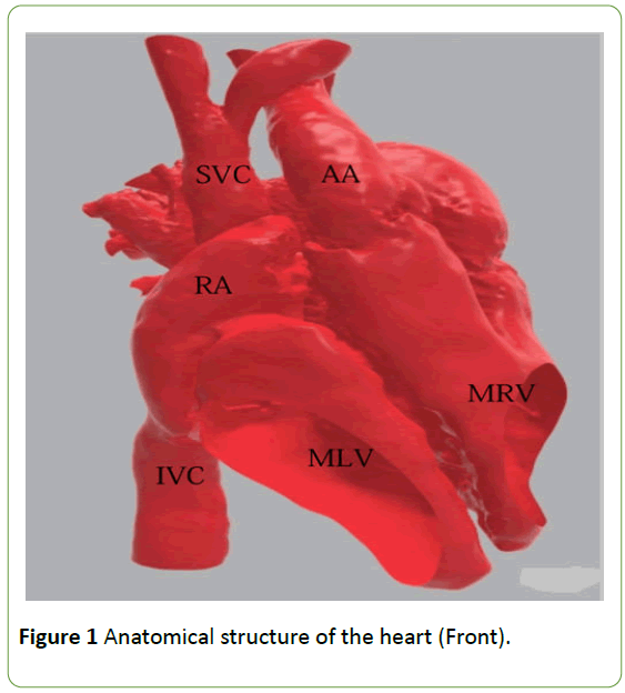
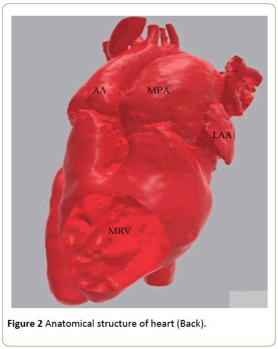
Because of the dangerous pulmonary resistance, we used oral Bosentan and Tadalafil combined to relieve pulmonary arterial pressure. And three months later, her cardiac catheterization showed the pulmonary resistance was significantly decreased to 9.20 Wood before oxygen inhalation and 2.93 Wood after oxygen inhalation. We use computed tomography to reconstruct the anatomical structure of the heart and established a 3D printed model (Figures 1 and 2) [3], with which we could accurately assure the complex malformation anatomies in the heart of the patient and develop a precise operation plan.
The operation was performed in a hypothermic cardiopulmonary bypass, using a median sternotomy. The ascending aorta and the superior and inferior vena cava were cannulated. After occluding the aorta, the right atrium was cut, the left-heart venting catheter was inserted through the atrial septum incision.
It was found that, the pulmonary artery was located to the left side and posterior to the aorta. The aorta and the pulmonary artery lose their cross connection and were arranged in parallel. Both great arteries were completely from the morphological right ventricle. The aorta and the pulmonary arteries were obviously thickened. Discordant atrioventricular connection was observed: morphologic left ventricle was to the right, the morphologic right ventricle was lying to the left of the morphologic left ventricle, with the blood in right-sided left ventricle passing through the ventricular septal defect to left-sided right ventricle. The blood of the two sides of the ventricles was mixed in anatomic right ventricle. The left ventricle was located on the right side and communicated with the right atrium through the mitral valve.
The right ventricle lay on the left side and communicated with the left atrium through the tricuspid valve. The direction of blood flow was from the left-sided tricuspid valve to pulmonary artery and aorta. The tricuspid annulus was larger than normal one and with mild tricuspid regurgitation. The biventricular development was balanced. The atrial septum was continuous and complete, and the diameter of peri membranous ventricular septal defect was about 1.50 cm × 1.80 cm. The ventricular septal defect was far away from the two great arteries and relatively close to the aorta. The aorta and the pulmonary artery had their own conus respectively. There was no stenosis under the aortic valve and the annulus, and no arterial duct was found.
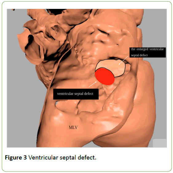
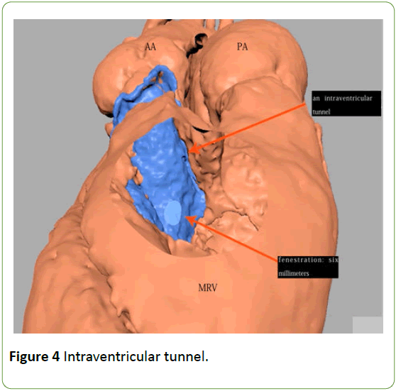
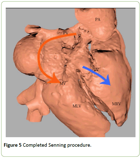
Proper resection of the partial hypertrophy of the right ventricular outflow tract was performed. Because it was restrictive to flow into a tunnel from the left ventricle, the ventricular septal defect was enlarged carefully, and a corresponding size 18 artificial vascular patch was placed. An intraventricular tunnel conducting the left ventricle blood flow to aorta was established using continuous and interrupted 5/0 prolene sutures. The reconstructed inner tunnel could pass the 18 #probe, keeping a small hole about 6 mm in diameter on this pipe (Figures 3 and 4). DeVega annuloplasty was accomplished by narrowing the anulus only in the lateral half of the anterior leaflet and the base of the posterior leaflet with the 4/0 prolene suture. After that, the Senning procedure was completed (Figure 5).
After de-airing and removal of the aortic clamp, the heart started to beat automatically, and the electrocardiograph indicated an atrioventricular block; the ventricular rate is 65 times per minute, and then an epicardial atrioventricular sequence temporary pacing wire was placed. The intraoperative transesophageal echocardiography showed that the connection of anatomy was normal, and the blood flow of the artificial pipe between the left ventricle and the aorta was unobstructed. The time of cardiopulmonary bypass was 205 minutes and the interruption time was 137 minutes.
Peri-operative pulmonary hypertension is an independent risk factor to influence prognosis. In the present case, the targeted therapeutic drug Remodulin and oral Bosentan and Tadalafil were used in combination to reduce pulmonary hypertension after operation [4,5]. At six months after the operation, the cardiac echocardiography showed that the heart function was good, except the systolic pressure of pulmonary artery was still high, and the ECG showed a complete atrioventricular block. The heart rate is maintained at 50 times per minutes. The patient reported no obvious discomfort. Concerning the long-term prognosis of the patient, we proposed to set up a pacemaker, which was not accepted by the patient’s family due to poor economic situation.
Discussion and Conclusion
There are various surgical procedures for correcting double outlet right ventricle with discordant atrioventricular connections, including Fontan procedure, intraventricular tunnel repair, the valved homograft conduit (such as Rastelli operation), REV procedure, and a variety of double-switch operations. Fontan procedure is suitable for palliative treatment of a ventricular dysplasia and intraventricular tunnel repair is more suitable for ventricular septal defect under the aortic valve or beneath the pulmonary artery, although the rationale of the intraventricular tunnel repair and the use of the valved conduit is still controversial. Our patient had no problem with the development of the main pulmonary artery, and the ventricular distance was relatively close to the aorta, and so we did not use the internationally popular double-switch or REV operation procedure to widen the right ventricle outflow tract.
In the literature reviewing the DORV with discordant atrioventricular connection, Dr Shin 'Oka has performed Senning or Mustrad plus Rastelli operations on 52 patients 2. It is reported that the right ventricle to pulmonary artery is connected by using the pericardial and other conduits. Rastelli procedure could significantly alleviate the stenosis of the right ventricle outflow tract. They have reported that 18 patients have superior ventricular tachycardia after operation because of atrial switch. The cause of complete atrioventricular block has been analyzed, suggesting that it is very easy to damage the conduction bundle when it is upward to enlarge the ventricular septal defect. Therefore, it's important to manage ventricular septal defect during operation [6]. Prophylactic epicardial pacing could reduce the incidence of complete atrioventricular block [7]. It is suggested that intraoperative electrophysiologic mapping should be used to reduce the damage of conduction tract, which may cause air embolus and has poor practicality [8]. The most effective way to prolong the survival period for patients with postoperative complete atrioventricular block is suggested to be implantation of an epicardial pacemaker.
Of note, Debin Liu has reported a case of a DORV (S.D.L) with sub-pulmonary ventricular septal defect [9]. They use arterial switch operation plus baffle from the ventricular septal defect to pulmonary artery. In our case, we didn't use arterial switch operation, based on the following considerations: (1) ventricular septal defect was close to the aorta and only a small operation was needed to establish the unobstructed relationship between the left ventricle and aorta; and (2) we needed to make the mitral and tricuspid valves adapt to the hemodynamic requirements of the systemic and pulmonary circulation, respectively, to achieve anatomical correction.
The DORV in the setting of discordant atrioventricular connection is an extremely rare disease. The report of treatment for older patients is scarce. In the present case, we used 3D printed technique in the operation. The postoperative complete atrioventricular block remains a concern for us. We will have a long-term follow-up on this patient and provide more valuable experience for clinical practice in the future.
Declarations
Ethical approval and consent to participate
The study was done after agreement from the local ethics committee and with the patients 'informed consent.
Consent for publication
Written informed consent was obtained from the patient for the publication of this case report and any accompanying images
Availability of supporting data
We confirm that the date has been read and approved by all named authors and that the date in this article comes from the results of the examination. We further confirm that the date is true, reliable and effective.
Competing interests
The authors declare that they have no competing interests.
Funding
The authors received no financial support for this research, author-ship, and publication of this article.
Author’s contributions
Wei designed the report and completed the manuscript. Peng, Zhang, and Ai oversaw patient operation. Hu and Zheng operated on the patient and were responsible for the postoperative care as well. Zhang and Ai revised the manuscript, and Peng supervised all the work. All the authors have read and approved the final manuscript.
Acknowledgements: The authors have no funding, financial relationships, or conflicts of interest to declare.
Authors' information
Wei is the postgraduate student in School of Medicine Henan University. Peng is my postgraduate tutor in Henan Provincial People’s Hospital, meanwhile, he is also the chief physician of children heart center ward 2. Ai, Zhang, Hu and Zheng are the surgeons of children heart center ward 2 in Fu Wai Central China Cardiovascular Hospital and Fu Wai Central China Cardiovascular Hospital is the heart center of Henan Provincial People’s Hospital.
References
- Battistessa S, Soto B (1990) Double outlet right ventricle with discordant atrioventricular connexion: An angiographic analysis of 19 cases. Int J Cardiol 27: 253-263.
- Shin'oka T, Kurosawa H, Imai Y, Aoki M, Ishiyama M, et al. (2007) Outcomes of definitive surgical repair for congenitally corrected transposition of the great arteries or double outlet right ventricle with discordant atrioventricular connections: risk analyses in 189 patients. J Thorac Cardiovasc Surg 133: 1318-1328.
- Dydynski PB, Kiper C, Kozik D, Keller BB, Austin E, et al. (2016) Three-dimensional reconstruction of intracardiac anatomy using cta and surgical planning for double outlet right ventricle: early experience at a tertiary care congenital heart center. World J Pediatr Congenit Heart Surg 7: 467-74.
- Skoro-Sajer N, Gerges C, Balint OH, Kohalmi D, Kaldararova M, et al. (2018) Subcutaneous treprostinil in congenital heart disease-related pulmonary arterial hypertension. Heart 2018.
- Iversen K, Jensen AS, Jensen TV, Vejlstrup NG, Sondergaard L (2010) Combination therapy with bosentan and sildenafil in Eisenmenger syndrome: a randomized, placebo-controlled, double-blinded trial. Eur Heart J 31: 1124-1131.
- Mavroudis C, Stewart RD, Prieto LR, Zahka K, Jacobs ML (2011) Management of the ventricular septal defect during double switch operation for atrioventricular discordant connections. Semin Thorac Cardiovasc Surg Pediatr Card Surg Annu 14: 29-34.
- Cohen MI, Rhodes LA, Spray TL, Gaynor JW (2004) Efficacy of prophylactic epicardial pacing leads in children and young adults. Ann Thorac Surg 78: 197-202.
- Ostermeyer J, Breithardt G, Kolvenbach R, Korfer R, Seipel L (1979) Intraoperative electrophysiologic mapping during cardiac surgery. Thorac Cardiovasc Surg 27: 260-70.
- Liu D, Chen C, Song B (2013) Arterial switch operation for double outlet right ventricle (S, D, L) with sub-pulmonary ventricular septal defect. Thorac Cardiovasc Surg 61: 312-315.

Open Access Journals
- Aquaculture & Veterinary Science
- Chemistry & Chemical Sciences
- Clinical Sciences
- Engineering
- General Science
- Genetics & Molecular Biology
- Health Care & Nursing
- Immunology & Microbiology
- Materials Science
- Mathematics & Physics
- Medical Sciences
- Neurology & Psychiatry
- Oncology & Cancer Science
- Pharmaceutical Sciences
