A Rare Case of Inguinal Hernia with Complete Appendix Herniation to the Scrotum
Farshad Banouei1* and Mehdi Komaki2
1Urology and Nephrology Research Center, Hamedan, Iran
2Department of Urology, Hamedan University of Medical Sciences, Hamedan, Iran
- *Corresponding Author:
- Farshad Banouei
Urology and Nephrology Research Center, Hamedan,
Iran,
E-mail: farshadbanouei90@gmail.com
Received date: February 27, 2023, Manuscript No. IPMCRS-23-16153; Editor assigned date: March 01, 2023, PreQC No. IPMCRS-23-16153 (PQ); Reviewed date: March 14, 2023, QC No. IPMCRS-23-16153; Revised date: March 21, 2023, Manuscript No. IPMCRS-23-16153 (R); Published date: March 28, 2023, DOI: 10.36648/2471-8041.9.3.268
Citation: Banouei F, Komaki M (2023) A Rare Case of Inguinal Hernia with Complete Appendix Herniation to the Scrotum. Med Case Rep Vol.9 No. 3:268.
Abstract
Introduction: Now days, inguinal hernia is one of the most common problems in different societies and at different ages. Despite pre-surgery investigations, in many cases, the contents of the hernia sac and the size of the defect are detected during surgery.
Case Presentation: Patient, an 83-year-old man, complained of inguinal swelling and right hemiscrotum. During surgery after the release of the cord, a worm-shaped appendage with a length of 23 cm was seen attached to the cord, which entered the scrotum parallel to it and it was inflamed and edematous, which was carefully separated and after complete release, it was connected to the cecum proximally, which was found to be an appendix.
Discussion: The contents of the sac of these hernias are very different and varied, of which the appendix is one of the rarest. Despite the diagnostic measures that are performed before surgery, in most cases, this form of hernia is diagnosed during surgery. In most cases, it is possible to act based on Losanoff and Basson's classification.
Conclusion: The presence of strong clinical suspicion by emergency doctors is very important in these cases. During herniorrhaphy surgery, the surgeon must be prepared to deal with this type of hernia.
Keywords
Appendix; Hernia; Surgery; Scrotum
Introduction
Nowadays, inguinal hernia is one of the most common problems in different societies and at different ages. The clinical manifestations of this disorder vary from asymptomatic to very bulky and painful cases and depending on the severity of symptoms and clinical manifestations, different treatment methods are used to treat it.
Inguinal hernia is divided into direct and indirect type, which is based on the location of the defect in the inguinal canal. The contents of the hernia sac also include a wide range of intraabdominal organs. Despite pre-surgery investigations, in many cases, the contents of the hernia sac and the size of the defect are detected during surgery. Among the organs that herniate into the inguinal region, the appendix is one of the rarest organs, so that in some studies, the prevalence was about 1% [1]. This condition is rare and the surgeon must be prepared to treat it and perform pre-surgery procedures.
Case Presentation
The patient, an 83-year-old man, complained of inguinal swelling and right hemiscrotum since about 10 years ago, which was accompanied by severe pain, swelling, and redness in the inguinal area and right hemiscrotum since about 2 weeks ago. The swollen area increased in size during the last 2 weeks, but no evidence of purulent discharge was seen. The patient did not have fever and did not complain of anorexia or nausea and vomiting. He had no urinary or digestive symptoms. The patient did not mention the history of any particular disease. In the examination, slight swelling of the inguinal area and the right hemiscrotum was seen and evidence of direct inguinal hernia was evident on examination and the hernia sac was easily removed. The abdomen was soft and had no tenderness or guarding.
In the ultrasound performed for the patient, a defect with a diameter of 12 mm was seen in the floor of the inguinal canal and there was herniation of fatty tissue into the inguinal canal, but no signs of intestinal tissue herniation into the inguinal canal were seen with or without Valsalva maneuver. In the scrotal area, fluid accumulation was seen in the right hemiscrotum along with the internal septa that destroyed the hydrocele. All tests performed on the patient were also normal.
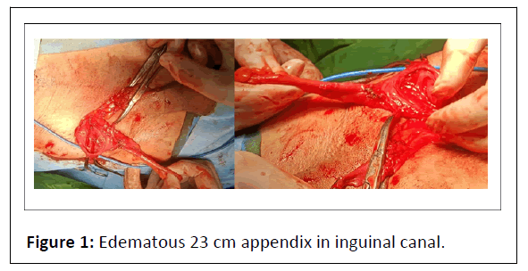
The patient was diagnosed with an inguinal hernia and was transferred to the operating room for repair. Under spinal anesthesia, in the supine position and after prep and drape, the skin and fascia were opened with a right inguinal incision and the spermatic cord was carefully released. After the release of the cord, a worm-shaped appendage with a length of 23 cm was seen attached to the cord, which entered the scrotum parallel to it and it was inflamed and edematous, which was carefully separated and after complete release, it was connected to the cecum proximally, which was found to be an appendix. The mentioned appendix was swollen and edematous (Figure 1).
Therefore, an appendectomy was performed and the liquid inside the scrotum, which was a reactive hydrocele, was drained and a drain was inserted. Then the defect of the inguinal canal floor was also repaired and the fascia and skin were repaired and the patient was transferred to recovery. The patient was hospitalized for 2 days and was discharged in good general condition and in the follow-up 2 weeks later, the symptoms were completely resolved and the surgical wound was completely healed.
Results and Discussion
There are different types of hernia; inguinal hernia is one of them [2]. The contents of the sac of these hernias are very different and varied, of which the appendix is one of the rarest and the presence of inflammation in the herniated appendix into the inguinal canal is very rare and has a prevalence of about 0.1% [3,4]. In most cases, the cause of inflammation and edema in the herniated appendix into the inguinal canal is the pressure inside the canal and the deep ring on the appendix and the disruption in blood supply and the exacerbation of infection in these cases [4]. This disorder was first discovered in 1736 at the George Hospital in an 11-year-old boy and the first appendectomy in the hernia sac was performed in this patient [5]. The presence of the appendix in the hernia sac, which is called Amyand's hernia, is more common in men than women [6]. This type of hernia usually occurs on the right side, but cases have also been seen on the left side as a result of displacement of the appendix due to the rotation of the intestine [7]. This type of hernia is divided into different types and depending on the severity of the inflammation and accompanying pathology and the treatment is carried out accordingly [8] (Table 1).
| Classification | Description | Surgical management |
|---|---|---|
| Type 1 | Normal appendix in an inguinal hernia | Hernia reduction, mesh repair |
| Type 2 | Acute appendicitis in an inguinal hernia, without abdominal sepsis | Appendectomy, primary repair of hernia without mesh |
| Type 3 | Acute appendicitis in an inguinal hernia, with abdominal wall or peritoneal sepsis | Laparotomy, appendectomy, primary repair without mesh |
| Type 4 | Acute appendicitis in an inguinal hernia, with abdominal pathology | Manage as type 1-3, investigate pathology as needed |
Table 1: Losanoff and Basson classification of Amyand’s hernia with their surgical management.
Despite the diagnostic measures that are performed before surgery, in most cases, this form of hernia is diagnosed during surgery.
In spite of the extensive studies that have been done on this type of hernia, a standard method that is generally agreed upon has not been approved and regarding the use of mesh in the repair of this type of hernia or prophylactic appendectomy (in cases diagnosed before the operation), there is a difference of opinion [9]. However, in most cases, it is possible to act based on Losanoff and Basson's classification [10,11].
Conclusion
The presence of appendix in the hernial sac is a rare disorder that, if not diagnosed in time and proper treatment measures, can cause inflammation and infection and cause serious problems for patients. Therefore, the presence of strong clinical suspicion by emergency doctors is very important in these cases.During herniorrhaphy surgery, the surgeon must be prepared to deal with this type of hernia.
References
- Patoulias D, Kalogirou M, Patoulias I (2018) Amyand’s hernia: An up-to-date review of the literature. Acta Medica 60: 131-4.
[Crossref], [Google Scholar], [Indexed]
- Namdev GH, Sanjay P, Varun S, Padnanabh D (2020) Amyand’s hernia: A case report. Int Surg J 7: 2072-2074.
[Crossref]
- Ikram S, Kaleem A, Ahmad S (2018) Amyand hernia: A literature review of the diagnosis and management of the rare presentation of the wandering appendix. J Rare Disord Diagn Ther 4: 1.
- Kathar hussain Mr, Kulasekeran N (2020) A case report of Amyand hernia-radiological diagnosis and literature review. EJRNM 51: 1-4.
[Crossref]
- Al Maksoud AM, Ahmed AS (2015) Left Amyand’s hernia: An unexpected finding during inguinal hernia surgery. Int J Surg Case Rep 14: 7-9.
[Crossref], [Google Scholar], [Indexed]
- Gurer A, Ozdogan M, Ozlem N, Yildirim A, Kulacoglu H, et al. (2006) Uncommon content in groin hernia sac. Hernia 10: 152-155.
[Crossref], [Google Scholar], [Indexed]
- Bhatti SI, Hashmi MU, Tariq U, Bhatti HI, Parkash J, et al. (2018) Amyand’s hernia: A rare surgical pathology of the appendix. Cureus 10: e2827.
[Crossref], [Google Scholar], [Indexed]
- Losanoff J, Basson M (2008) Amyand hernia: A classification to improve management. Hernia 12: 325-326.
[Crossref], [Google Scholar], [Indexed]
- Michalinos A, Moris D, Vernadakis S (2014) Amyand's hernia: A review. Am J Surg 207: 989-995.
[Crossref], [Google Scholar], [Indexed]
- Morales-Cárdenas A, Ploneda-Valencia CF, Sainz-Escárrega VH, Hernández-Campos AC, Navarro-Muniz E, et al. (2015) Amyand hernia: Case report and review of the literature. Ann Med Surg 4: 113-115.
[Crossref], [Google Scholar], [Indexed]
- Yagnik VD (2011) Amyand hernia with appendicitis. Clin Pract 1: e24.
[Crossref], [Google Scholar], [Indexed]

Open Access Journals
- Aquaculture & Veterinary Science
- Chemistry & Chemical Sciences
- Clinical Sciences
- Engineering
- General Science
- Genetics & Molecular Biology
- Health Care & Nursing
- Immunology & Microbiology
- Materials Science
- Mathematics & Physics
- Medical Sciences
- Neurology & Psychiatry
- Oncology & Cancer Science
- Pharmaceutical Sciences

