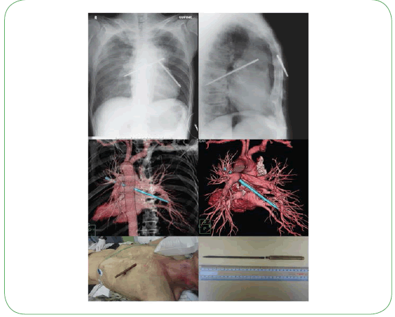
A Close Call: Lucky Man Survives Penetrating Chest Trauma
Michihisa Terado, Atsuyoshi Iida, Kohei Tsukahara, Keiji Sato, Masami Takagaki, Takahiro Hirayama and Atsunori Nakao
Michihisa Terado1, Atsuyoshi Iida1, Kohei Tsukahara1, Keiji Sato1, Masami Takagaki2, Takahiro Hirayama1 and Atsunori Nakao1*
1Department of Emergency and Critical Care Medicine, Okayama University, Graduate School of Medicine Dentistry and Pharmaceutical Sciences, Japan
2Cardiovascular Surgery, Okayama University, Graduate School of Medicine Dentistry and Pharmaceutical Sciences, Japan
- *Corresponding Author:
- Atsunori Nakao
Department of Emergency and Critical Care Medicine
Okayama University, Graduate School of Medicine
Dentistry and Pharmaceutical Sciences
Okayama, Japan
Tel: +81-86-235-7426
E-mail: qq-nakao@okayama-u.ac.jp
Received date: April 16, 2016; Accepted date: April 18, 2016; Published date: April 22, 2016
Citation: Terado M, Iida A, Tsukahara K, et al. A Close Call: Lucky Man Survives Penetrating Chest Trauma. Med Case Rep. 2016, 2:2.
The survival of a patient with a thoracic impalement injury is an extremely rare event, especially if the impalement occurs on the left side of the chest. A 67-year old male attempted suicide by advancing a metal file into his right chest. His vital signs were stable and computed tomography demonstrated that the foreign object had broken into two pieces, missing vital internal structures including greater vessels (aorta or pulmonary artery) and reaching the right lung. The patient was taken immediately to the operating room for emergency surgery. Under cardiopulmonary bypass, the foreign body was removed and perforating wounds of the right atrium and peripheral branch of the pulmonary artery were repaired. His postoperative course was uneventful. Understanding the extent of such an injury is extremely important in order to plan the appropriate surgical approach. The impaling object must not be removed until reaching the hospital. Rapid transport of the patient to a qualified hospital is critical and emergency surgery is mandatory.
Abstract
The survival of a patient with a thoracic impalement injury is an extremely rare event, especially if the impalement occurs on the left side of the chest. A 67-year old male attempted suicide by advancing a metal file into his right chest. His vital signs were stable and computed tomography demonstrated that the foreign object had broken into two pieces, missing vital internal structures including greater vessels (aorta or pulmonary artery) and reaching the right lung. The patient was taken immediately to the operating room for emergency surgery. Under cardiopulmonary bypass, the foreign body was removed and perforating wounds of the right atrium and peripheral branch of the pulmonary artery were repaired. His postoperative course was uneventful. Understanding the extent of such an injury is extremely important in order to plan the appropriate surgical approach. The impaling object must not be removed until reaching the hospital. Rapid transport of the patient to a qualified hospital is critical and emergency surgery is mandatory.

Open Access Journals
- Aquaculture & Veterinary Science
- Chemistry & Chemical Sciences
- Clinical Sciences
- Engineering
- General Science
- Genetics & Molecular Biology
- Health Care & Nursing
- Immunology & Microbiology
- Materials Science
- Mathematics & Physics
- Medical Sciences
- Neurology & Psychiatry
- Oncology & Cancer Science
- Pharmaceutical Sciences

