A Case Report of Undiagnosed Pheochromocytoma
de Bie L*
Department of Medical, Gujranwala Institute of Nuclear Medicine and Radiotherapy, Gujranwala, Pakistan
- *Corresponding Author:
- De Bie L
Department of Medical,
Gujranwala Institute of Nuclear Medicine and Radiotherapy, Gujranwala,
Pakistan;
Email: lianobie@unira.it
Received date: September 26, 2022, Manuscript No. IPMCRS-22-14635; Editor assigned date: September 29, 2022, PreQC No. IPMCRS-22-14635 (PQ); Reviewed date: October 14, 2022, QC No. IPMCRS-22-14635; Revised date: February 10, 2023, Manuscript No. IPMCRS-22-14635 (R); Published date: February 17, 2023, DOI: 10.36648/2471-8041.9.2.263
Citation: de Bie L (2023) A Case Report of Undiagnosed Pheochromocytoma. Med Case Rep Vol:9 No:2
Abstract
Giant Cystic Pheochromocytomas (GPCCs) are rare adrenal tumors and the majority of them present as asymptomatic. As a result GPCCs often remain undiagnosed until surgery and therefore the surgical teams face a greater challenge in perioperative management. The present study describes the case of a 36 year‑old woman with an undiagnosed GPCC, which was successfully resected despite the occurrence of perioperative cardiovascular events, including hypertension, hypotension, ventricular arrhythmias, acute heart failure, acute myocardial infarction and the patient was discharged home without any recurrence. It should be considered in retroperitoneal tumour of patients with nonspecific symptoms and given adequate treatment to promote the perioperative safety.
Keywords
Lipomyelomeningocele; Aponeurosis Osteotomy; Tibialis anterior muscle; Fibulamuscle
Introduction
Equinovarus is one of the most common malformations in the motor system and adult rigid equinovarus is the most difficult category of surgical orthotics and functional reconstruction [1]. Equinovarus occurrence reason is caused by innate or acquired a variety of factors, congenital lesions in fetal period of primary development is mainly due to the limb, evolved into the Equinovarus deformity, the day after tomorrow is mainly due to the nervus peroneus communis injury sequela, foot ankle trauma sequela, stroke and cerebral hemorrhage sequelae, lesions caused by polio. This case is a congenital Equinovarus deformity due to lipomyelomeningocele,which is reported as follows.
Case Presentation
A 20-year-old male patient was diagnosed with congenital neural tube malformation-Lipomyelomeningocele (LMM) 10 years ago when he was found to have bipedal varus and decreased muscle strength [2]. He underwent spinal cord surgery at that time. Six years after the operation, the left foot varus was significantly worsened, the left ankle flexion and extension was limited, the ankle joint plantar flexion was 70â??, and the average range of motion was 10â?? (plantar flexion 10â??-back extension 0°); tibialis anterior muscle and fibulamuscle strength level 0, tibialis posterior muscle strength level 2, raise the medial longitudinal arch, plantar ulceration, sphincter bladder function paralysis, and catheterization bag for 5 years [3].
Use general anesthesia, patient in supine position. First, a small longitudinal incision was made on the medial side of the Achilles tendon, and a sharp knife was used to cut the tendon vertically [4]. The Achilles tendon was released in the shape of "Z". After proper pulling, the arteries and veins and nerves of the posterior tibial were separated for protection, and the soft tissue and the posterior tibial joint capsule were released [5]. The insertion point of the posterior tibial tendon was cut through the medial foot incision, and the tendon was extracted before the ankle to prepare for the external displacement of the posterior tibial tendon. The plantar medial incision was made to expose the plantar aponeurosis, and "Z" was released to suture the skin.
A 36-year-old woman presented to Qilu Hospital of Shandong University on May 9, 2013, with the primary complaint of abdominal discomfort following eating and lumbodorsal distending pain for 3 months and reported weight loss of 8 kg during this time. The patient's medical history included a caesarean section and an ovarian cysts surgery, but no history of hypertension or headache. The patient's vital signs included an arterial blood pressure of 120/80 mmHg, heart rate of 80 bpm and temperature 36.8°C. The only significant finding during physical examination was for left renal region percussion pain. Laboratory analysis identified a slightly elevated blood glucose level of 7.73 mmol/l (normal range, 3.90 mmol/l-6.10 mmol/l).
Results
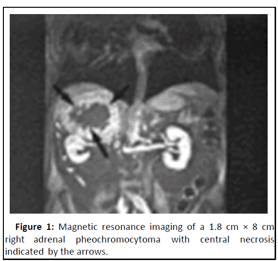
Ultrasonography examination revealed a cystic spaceoccupying lesion (10.3 cm × 9.3 cm) in the left upper abdomen, which was considered to be a left renal cystic mass. Abdominal Computed Tomography (CT) and a contrast-enhanced CT scan demonstrated a giant cystic-solid mass on the left kidney, which occupied a large part of the superior abdominal cavity (Figure 1). Based on the patient's age, gender, history, physical examination and preoperative imaging, a clinical diagnosis of a malignantadrenal mass was suspected. No special treatment was given prior to surgery because the diagnosis of a pheochromocytoma had not been considered [6].
Discussion
The dorsal incision of the first metatarsal was made, the skin was cut subcutaneously, the first metatarsal base was exposed and the first metatarsal base was cuneate osteotomy, fixed with 6-hole "T" type plate and screw and the internal arch of the foot was reduced. Make an incision at the calcaneocuboid joint on the dorsolateral side of the foot, cut the skin subcutaneously, expose the calcaneocuboid joint, remove the cartilage surface, expose the fresh bone surface, "X" fix with steel plate and screw to correct the foot adduction. The lateral incision of the calcaneus was made, the skin was cut subcutaneously, the external side of the calcaneus was exposed, the bone knife was osteotomized and externally removed, and fixed with a 4.5 mm hollow screw to correct the calcaneal varus. Intraoperative fluoroscopy showed that the position of steel plate, screw and hollow screw was good and the length was appropriate [7]. The incision was made at the lateral calcaneus, the skin was cut subcutaneously, the peroneus brevis was separated, the tendons of the anterior ankle and the posterior tibial tendon were moved and sutured to strengthen the eversion force, the muscle balance was observed during the operation, the varus of the foot, high arch and plantarsal flexion were corrected, the wound was rinsed, the layer by layer was sutured and the back extension was fixed with plaster. The operation was successful, the anesthesia was satisfactory and the patient was returned to the ward after the operation.
Conclusion
A continuous infusion of phentolamine was used in a patient with pheochromocytoma to control perioperative hypertensive episodes during surgical adrenalectomy. The management of patients with pheochromocytoma remains a challenge for the anesthesiologist despite the advent of new drugs and techniques. Treating patients preoperatively with alphaadrenergic blockade is helpful for reducing intraoperative hypertensive episodes, thus decreasing morbidity and mortality.
References
- Myklejord DJ (2004) Undiagnosed pheochromocytoma: The anesthesiologist nightmare. Clin Med Res 2:59-62
[Crossref] [Google Scholar] [PubMed]
- Dabbous A, Siddik-Sayyid S, Baraka A (2007) Catastrophic hemodynamic changes in a patient with undiagnosed pheochromocytoma undergoing abdominal hysterectomy. Anesth Analg 104:223-224
[Crossref] [Google Scholar] [PubMed]
- Sarathi V, Lila AR, Bandgar TR, Menon PS, Shah NS (2010) Pheochromocytoma and pregnancy: A rare but dangerous combination. Endocr Pract 16:300-309
[Crossref] [Google Scholar] [PubMed]
- Bajwa SS, Bajwa SK (2011) Implications and considerations during pheochromocytoma resection: A challenge to the anesthesiologist. Indian J Endocrinol Metab 15:337-344
[Crossref] [Google Scholar] [PubMed]
- Sbardella E, Grossman AB (2020) Pheochromocytoma: An approach to diagnosis. Best Pract Res Clin Endocrinol Metab 34:101346
[Crossref] [Google Scholar] [PubMed]
- Dugas G, Fuller J, Singh S, Watson J (2004) Pheochromocytoma and pregnancy: A case report and review of anesthetic management. Can J Anaesth 51:134-138
[Crossref] [Google Scholar] [PubMed]
- Kuok CH, Yen CR, Huang CS, Ko YP, Tsai PS (2011) Cardiovascular collapse after labetalol for hypertensive crisis in an undiagnosed pheochromocytoma during cesarean section. Acta Anaesthesiol Taiwan 49:69-71
[Crossref] [Google Scholar] [PubMed]

Open Access Journals
- Aquaculture & Veterinary Science
- Chemistry & Chemical Sciences
- Clinical Sciences
- Engineering
- General Science
- Genetics & Molecular Biology
- Health Care & Nursing
- Immunology & Microbiology
- Materials Science
- Mathematics & Physics
- Medical Sciences
- Neurology & Psychiatry
- Oncology & Cancer Science
- Pharmaceutical Sciences

