Abstract
Desoxycorticosterone (DOC) adrenal normotensive adrenal tumor: A rare case
A 31 year-old man was admitted with low back pain (LBP).He had LBP since four months ago. He also suffered from severe weakness since one year ago. He had anorexia and about 8 kilogram weight loss during last year. He also complained of polydipsia and polyuria. Physical examination revealed tenderness on L2 vertebrae and loss of dorsiflexion in left foot and positive left SLR. Physical exam was normal otherwise. Vital signs were normal and blood pressure was 110/70. Hypokalemia ranged from 1.4-3mg/dl was detected. 24 hour urinary volume was 3850cc and urine specific gravity was 1006. 24 hour urinary potassium concentration was 54meq/L (normal range=25-125). Lateral lumbosacral radiography showed decreased bone density, intervertebral space and vertebral height. These findings did not signify the low back pain. CT scan of lumbosacral vertebras showed lithic destructive lesion at body of L2 accompanied with L1-L2 intervertebral disc involvement and paravertebral abscess. Metastatic bone lesion was the radiologic diagnosis. In order to confirm the nature of the lesion open biopsy was done. Metastatic adenocarcinoma was the anatomical diagnosis. Abdominal ultrasonography showed a70x150 millimeter lobulated mass with irregular border in retroperitoneal space next to spine with left kidney invasion. Abdominal and pelvic CT scan showed left adrenal mass with several calcification focuses and pesoas muscle involvement. Other organs were normal. Hypokalemia had induced nephrogenic diabetes insipidus and polyuria and polydipsia. For evaluation of hypokalemia several laboratory tests were conducted. Aldosterone was low and plasma rennin activity was low. Other hormonal evaluations were normal. According to laboratory data’s mineralocorticoid excess other than aldosterone was considered and deoxycorticosterone level was measured which was high (4.3ng/ml). After correction of hypokalemia, surgery proceeded and adrenal mass removed. The pathologic exam revealed a 11x7x5 centimeter mass with large necrotic and hemorrhagic focuses and cells with vesiculated, nucleolated nucleous accompanied with numerous mitotic and also atypical mitosis in some foci and granular eosinophilic cytoplasm, indicating adrenal carcinoma. According to Weiss system, pathologic staging was IV. Hypokalemia resolved after surgery. The patient was referred for radiation treatment of vertebrae after he was discharged. After radiation, adjuvant treatment with Mitotan 2g /day was instituted and no recurrence was observed up to 24 months follow up.
Author(s):
Shokoufeh Bonakdaran
Abstract | Full-Text | PDF
Share this

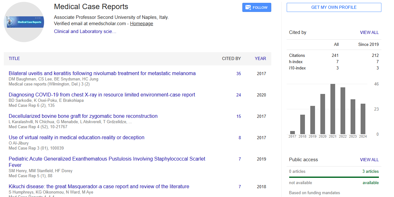
Google scholar citation report
Citations : 241
Medical Case Reports received 241 citations as per google scholar report
Medical Case Reports peer review process verified at publons
Abstracted/Indexed in
- Google Scholar
- China National Knowledge Infrastructure (CNKI)
- Cosmos IF
- Directory of Research Journal Indexing (DRJI)
- WorldCat
- Publons
- Secret Search Engine Labs
- Euro Pub


Open Access Journals
- Aquaculture & Veterinary Science
- Chemistry & Chemical Sciences
- Clinical Sciences
- Engineering
- General Science
- Genetics & Molecular Biology
- Health Care & Nursing
- Immunology & Microbiology
- Materials Science
- Mathematics & Physics
- Medical Sciences
- Neurology & Psychiatry
- Oncology & Cancer Science
- Pharmaceutical Sciences