Sonographic Non-Visualization of the Gallbladder: A Pitfall in Diagnosis of Emphysematous Cholecystitis
Zaw Min
DOI10.21767/2471-8041.100041
Zaw Min*
Division of Infectious Diseases, Allegheny General Hospital, Allegheny Health Network, Pittsburgh, PA, USA
- *Corresponding Author:
- Zaw Min
Division of Infectious Diseases
Allegheny General Hospital
Allegheny Health Network, 420 East North
Avenue, East Wing, Suite 407
Pittsburgh, PA 15212, USA
Tel: +1-412-359-3683
Fax: +1-412-359-3373
E-mail: zaw.min@ahn.org
Received date: December 05, 2016; Accepted date: March 20, 2017; Published date: March 22, 2017
Citation: Min Z. Sonographic Non-Visualization of the Gallbladder: A Pitfall in Diagnosis of Emphysematous Cholecystitis. Med Cas Rep. 2017, 3:1. doi:10.21767/2471-8041.100005
Abstract
Emphysematous cholecystitis is an uncommon complication of calculous cholecystitis due to secondary infection of gallbladder wall with gas-forming enteric organisms. It carries high morbidity and mortality to the patients and a rapid and early diagnosis is essential in patient’s clinical outcomes. Ultrasonography is usually the first and foremost test performed in cases of suspected biliary diseases. Herein, we describe a diagnostic pitfall of ultrasound in a patient with emphysematous cholecystitis – non-visualization of gallbladder, caused by reverberations from air in the gallbladder wall.
Keywords
Emphysematous cholecystitis; Nonvisualization of gallbladder; Sonographic findings
Introduction
Emphysematous cholecystitis is rare clinical sequelae of acute cholecystitis. A high mortality (15%) was documented in cases of emphysematous cholecystitis [1]. Although computed tomography is the imaging diagnosis of choice, ultrasound is almost always the first initial test ordered. Sonographic finding, highly suggestive of emphysematous cholecysitis, is known as the effervescent gallbladder in which multiple echogenic foci rising from the bottom to the top within the gallbladder resembling champagne gas bubbles [2].
However, a high clinical index of suspicion of emphysematous cholecystitis should be raised in an appropriate clinical setting if the ultrasound reported “nonvisualization” or “obscured” gallbladder because the entire gallbladder wall could be filled with gas, obscuring visualization of the gallbladder. In the present case, we highlight this important sonographic finding which should be regarded as one of the ultrasound spectrums of emphysematous cholecystitis.
Case Report
A 73-year-old man presented with fever, lethargy and malaise. His past medical history was significant for diabetes mellitus, cholelithiasis, sick sinus syndrome on pacemaker, and end-stage renal disease with hemodialysis via the right radiocephalic arteriovenous fistula. Physical examination showed temperature of 101°F, anicteric sclerae and tenderness over the right upper quadrant abdomen. Murphy’s sign was negative.
Pacemaker pocket site did not show evidence of fluid collection or overlying cellulitis. Significant laboratory investigations included leukocytosis of 11,000 cells/mm3 (normal 4000-10,000 cells/mm3) and normal liver function panel. Empiric therapy with intravenous vancomycin and piperacillin-tazobactam was initiated. The patient continued to be feverish (maximum temperature of 102.6°F) with intermittent pain over the right upper quadrant abdomen while receiving broad-spectrum antibiotics.
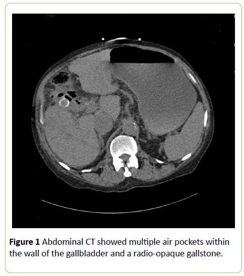
Blood cultures did not grow microorganisms. Ultrasonography of abdomen did not elicit sonographic Murphy’s sign; the sonographer failed to localize the gallbladder and reported as “non-visualization of gall-bladder”. Given persistent fever and abdomen pain, computed tomography (CT) of abdomen was performed. It demonstrated foci of air pockets within the gallbladder wall and a gallstone (Figure 1), suggestive of emphysematous cholecystitis. The patient underwent emergency open cholecystectomy and debridement of necrotic gallbladder tissues.
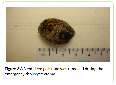
An oblong-shaped gallstone was extracted (Figure 2). Necrotic gallbladder tissue culture did not isolate microorganisms, however. The patient had an uneventful recovery and was discharged home with intravenous piperacillin-tazobactam for 2 weeks.
Discussion
Acute emphysematous cholecystitis is a rare infectious complication of acute cholecystitis. It is characterized by presence of gas within the lumen and wall of the gallbladder by gas-forming microorgansims [3]. It has a distinct male preponderance in contrast to that of the usual type of acute cholecystitis. Affected patients are usually middle-aged to elderly individuals (age between 50-70 years). Common risk factors include patients with diabetes mellitus and presence of gallstones [1,4]. Its pathogenesis is considered to be different from that of uncomplicated acute cholecystitis. Among others, the widely accepted pathogenic mechanism of emphysematous cholecystitis is thought to be compromise of vascular supply to the gallbladder, followed by gallbladder necrosis and secondary infection with gas-producing bacteria mainly Clostridium perfringens, often together with other gut flora such as Escherichia coli, Klebsiella pneumoniae, Pseudomonas aeruginosa, or anaerobic streptococci [1-4].
Diagnosis of acute emphysematous cholecystitis is challenging because clinical presentations are indistinguishable from those of uncomplicated acute cholecystitis. Ultrasonography is considered to be the initial test of choice for the diagnosis of hepatobiliary pathology. The various ultrasound features of emphysematous cholecystitis depend on the amount and location of gas within the lumen or wall of the gallbladder [4,5]. One of its sonographic characteristic findings is described as the “effervescent gallbladder” with floating multiple echogenic foci in the lumen of the gallbladder like champagne bubbles [2]. When a layer of the gas within the gallbladder wall is circled around the gallbladder, it usually obscures the visualization of gallbladder on the ultrasound, resulting in reporting of “non-visualized gallbladder”, which could easily be disregarded by clinicians. In this case, computed tomography is very helpful and becomes a diagnostic modality of choice for complicated cholecystitis [4-6]. Surgical therapy is always the mainstay of management of emphysematous cholecystitis with adjunctive antimicrobial treatment. Mortality of emphysematous cholecysitis is significantly higher (15%) than that of acute uncomplicated cholecystitis (4%) due to its life-threatening complications, such as gallbladder gangrene, perforation, abscess, and bile peritonitis [1]. Hence, while ultrasound report documents visualization of the gallbladder is obscured by gas, a high index of clinical suspicion is required for diagnosis of emphysematous cholecystitis in appropriate clinical context.
Conclusion
In conclusion, it is important for practicing clinicians and interpreting radiologists to recognize the ultra-sonographic pitfall of diagnosing acute emphysematous cholecystitis. “Nonvisualized gallbladder” is considered to be part of the spectrum of sonographic findings of emphysematous cholecystitis. Prompt diagnosis of emphysematous cholecystitis is critical because a delayed diagnosis would have a major impact on the patient’s clinical outcome.
References
- Bernstein D, Soeffing J, Daoud YJ, Fradin J, Kravet SJ (2007) The obscured gallbladder. Am J Med 120:675-677.
- Nemcek AA Jr, Gore RM, Vogelzang RL, Grant M (1988) The effervescent gallbladder: A sonographic sign of emphysematous cholecystitis. Am J Roentgenol 50: 575-577.
- Sherlock S, Dooley J (2002) Gallstones and inflammatory gallbladder diseases. (11edn) Diseases of the liver and biliary system. London: Blackwell Scientific, UK, p: 610-612.
- Konno K, Ishida H, Naganuma H, Sato M, Komatsuda T, et al. (2002) Emphysematous cholecystitis: sonographic findings. Abdom Imaging 27: 191-195.
- Bloom RA, Libson E, Lebensart PD, Craciun E, Klin B, et al. (1989) The ultrasound spectrum of emphysematous cholecystitis. J Clin Ultrasound 17: 251-256.
- Charalel RA, Jeffrey RB, Shin LK (2011) Complicated cholecystitis: The complementary roles of sonography and computed tomography. Ultrasound Q 27: 161-170.

Open Access Journals
- Aquaculture & Veterinary Science
- Chemistry & Chemical Sciences
- Clinical Sciences
- Engineering
- General Science
- Genetics & Molecular Biology
- Health Care & Nursing
- Immunology & Microbiology
- Materials Science
- Mathematics & Physics
- Medical Sciences
- Neurology & Psychiatry
- Oncology & Cancer Science
- Pharmaceutical Sciences


