The Right Middle and the Inferior Pulmonary Vein Forming a Common Trunk
Yuriko Terada and Yoshiaki Furuhata
DOI10.21767/2471-8041.100058
Yuriko Terada* and Yoshiaki Furuhata
Department of Thoracic Surgery, Japanese Red Cross Medical Center, Tokyo, Japan
- *Corresponding Author:
- Yuriko Terada
Clinical Fellow, Department of Thoracic Surgery
Japanese Red Cross Medical Center, 4-1-22 Hiroo
Shibuya-Ku, Tokyo 113-8655, Japan
Tel: +81-3-3400-1311
Fax: +81-3-3409-1604
E-mail: yuriko.terada@gmail.com
Received Date: May 04, 2016; Accepted Date: May 10, 2017; Published Date: May 12, 2017
Citation: Terada Y, Furuhata Y. The Right Middle and the Inferior Pulmonary Vein Forming a Common Trunk. Med Case Rep, 2017, 3:3. DOI: 10.21767/2471-8041.100058
Abstract
We herein report a rare surgical case of the right middle lobe vein draining into the inferior pulmonary vein. The anatomical abnormalities in pulmonary veins can have a serious impact on complications during pulmonary surgery. Surgeons must be aware of any variation in the pulmonary vessels, and preoperative assessment of pulmonary venous anomalies is important during pulmonary surgery.
Keywords
Lung cancer; Pulmonary vein; Anatomical anomaly; Common trunk
Abbreviations
RMV: Right Middle Lobe Vein; IPV: Inferior Pulmonary Vein; CT: Computed Tomography
Case Presentation
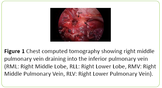
We herein report a rare surgical case of the right middle lobe vein (RMV) draining into the inferior pulmonary vein (IPV). A 49-year-old woman was incidentally found to have a 15 mm pure ground glass nodule in the right lower lobe on chest computed tomography (CT). Primary lung cancer with a clinical stage of T1aN0M0 stage IA was suspected. Thoracoscopic right lower wedge resection was performed, followed by right lower lobectomy and lymph node dissection. During surgery, it was observed that the RMV emptied into the inferior pulmonary vein (Figure 1).
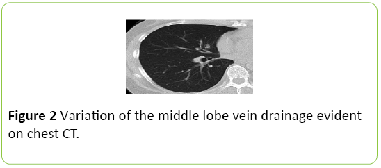
After the anterior part of the major fissure was divided, we carefully ligated and divided the inferior pulmonary vein using a vascular stapling device without injuring the middle pulmonary vein. Pathological examination of the resected specimen resulted in a diagnosis of T1aN1M0 stage IIB adenocarcinoma. Retrospectively, variation of the middle lobe vein drainage was evident on chest CT (Figure 2).
Discussion
Normally, the middle lobe vein unites to form the superior pulmonary vein, whereas in this case, the right middle lobe vein drained into the inferior pulmonary vein. Variations of pulmonary veins have been reported [1,2]. If these abnormalities are not considered, life-threatening complications such as uncontrolled bleeding, severe lung edema, or a greater extent of lung resection than expected may occur [3,4]. The efficacy of preoperative assessment of pulmonary vessels has been reported using three-dimensional CT for better understanding of the anatomical structure [5].
Conclusion
We highlight that surgeons must be aware of any variation in the pulmonary vessels, and preoperative assessment of pulmonary venous anomalies is important during pulmonary surgery, especially lobectomy.
References
- Cronin P, Kelly AM, Desjardins B, Patel S, Gross BH, et al. (2007) Normative analysis of pulmonary vein drainage patterns on multidetector CT with measurements of pulmonary vein ostial diameter and distance to first bifurcation. AcadRadiol 14: 178-188.
- Subotich D, Mandarich D, Milisavljevich M, Filipovich B, Nikolich V (2009) Variations of pulmonary vessels: some practical implications for lung resections. ClinAnat 22: 698-705.
- Sugimoto S, Izumiyama O, Yamashita A, Baba M, Hasegawa T (1998) Anatomy of inferior pulmonary vein should be clarified in lower lobectomy. Ann ThoracSurg 66: 1799-1800.
- Matsumoto I, Ohta Y, Tsunezuka Y, Sawa S, Fujii S, et al. (2005) A surgical case of lung cancer in a patient with the left superior and inferior pulmonary veins forming a common trunk. Ann ThoracCardiovascSurg 11: 316-319.
- Akiba T, Marushima H, Odaka M, Harada J, Kobayashi S, et al. (2010) Pulmonary vein analysis using three-dimensional computed tomography angiography for thoracic surgery. Gen ThoracCardiovascSurg 58: 331-335.

Open Access Journals
- Aquaculture & Veterinary Science
- Chemistry & Chemical Sciences
- Clinical Sciences
- Engineering
- General Science
- Genetics & Molecular Biology
- Health Care & Nursing
- Immunology & Microbiology
- Materials Science
- Mathematics & Physics
- Medical Sciences
- Neurology & Psychiatry
- Oncology & Cancer Science
- Pharmaceutical Sciences


