Lateral Condyle Temporary Hemiepiphysiodesis in Treatment of Cubitus Varus Deformity Post Supracondylar Humerus Fracture: A Case Report
Amnah Almahmudi*, Dhafer Alshehri, and Hussain Assaggaf
Department of Orthopaedic Surgery, East Jeddah Hospital, Saudi Arabia
- Corresponding Author:
- Amnah Almahmudi
Department of Orthopaedic Surgery
East Jeddah Hospital, Saudi Arabia
Tel: + 966122327555,
E-mail: amm99n@hotmail.com
Received date: June 24, 2020; Accepted date: August 04, 2020; Published date: August 11, 2020
Citation: Almahmudi A, Alshehri D, Assaggaf H (2020) Lateral Condyle Hemiepiphysiodesis in Treatment of Cubits Varus Deformity Post Supracondylar Humours Fracture: A Case Report. Med Case Rep Vol.6 No.4:150. DOI: 10.36648/2471-8041.6.4.150.
Abstract
A cubitus varus deformity often seen as a complication of a malunited supracondylar humerus fracture in children. several surgical management have been described to correct this deformity in this case report we described a Lateral condyle hemiepiphysiodesis as a surgical treatment in a 10 years old boy with a cubitus varus deformity post supracondylar humerus fracture with the outcome in 1 year follow up.
Keywords
Cubitus varus; Deformity; Hemiepiphysiodesis; Supracondylar humerus fracture
Introduction
Humerus Supracondylar fracture has been recognised since the time of Hippocrates as one of the common fractures in pediatric age group [1,2]. Gun stock or Cubitus varus deformity of the elbow is the most common complication following malunited supracondylar fracture of the humerus in skeletal immature children [3]. Various causes have been suggested, the frequent cause of cubitus varus deformity is malunited supracondylar humeral fragment rather than growth disturbance, Osteonecrosis either with or without growth arrest is considering a rare cause [4]. This deformity not only involving loss of the coronal alignment that make the forearm and hand deviate or adducted toward the midline of the body during elbow extension [5], but also has sagittal and axial planes deformity, summarized in recurvatum deformaty and internal rotation deformity respectively. Recurvatum deformity is in the plane of joint motion and remodels well. The internal rotation deformity is compensated by the shoulder movements and also tolerated well. Both of sagittal and axial deformities do not require any corrections and mostly the correction is focussed on the coronal plane deformity [1], although this deformity does not cause functional limitation [5], Cosmetic appearance is still the most common reason for presenting the parents with their child in the clinic [1].
Case Report
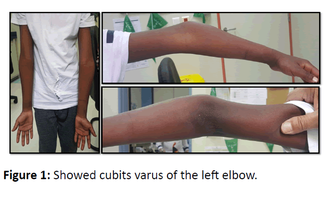
A 10-year-old boy presented with non-dominant left elbow cubitus varus deformity. According to the mother, the patient had diagnosed with a supracondylar humerus fracture five years ago in a local hospital after history of falling on outstretched hand, there he was treated conservatively on above elbow cast for a period of time. After removal of the cast, the parents noticed the deformity of the left elbow which was kept on progressing over the next five years. At presentation, there were no old records, so the exact type of fracture and the follow up was not known on clinical examination patient had a cubitus varus deformity of the left elbow (Figure 1) with painless full movement of the elbow comparing with the other side.
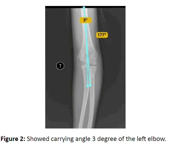
X-rays was done for the left elbow and showed reducing of normal carrying angle (Figure 2). As the deformity was progressive, his mother was told about the need of surgical intervention, informed consent was obtained for the surgery, and pre-operative planning was done for hemiepiphysiodesis of the lateral condyle as a day case surgery.
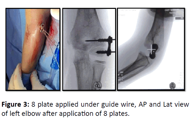
Patient was operated under general anesthesia, in supine position and prophylactic antibiotic was given. Tornequet wass applied as a standby, Drape and prep done of the left upper limb, around 1 cm longitudinal incision was done over the lateral condyle, 8 plates was applied under guide wire and carm, then fixed with 2 screws. Closure done after irrigation and sterile dressing applied and the limb was put to rest in arm sling (Figure 3).
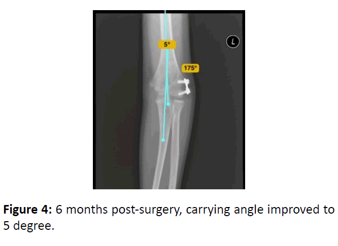
Patient discharged home afternoon, Exercise started once the patient pain free, clinical check-up and X-ray had performed during the follow up (Figure 4).
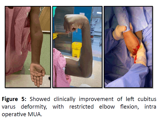
After 1 year follow up, the patient was having a corrected cubits varus deformity, with restricted elbow flexion with 100 degree due to missing of some sessions of physical therapy, so Patient booked for 8 plate removal and manipulation under anesthesia of the left elbow which was done as a day case surgery (Figure 5).
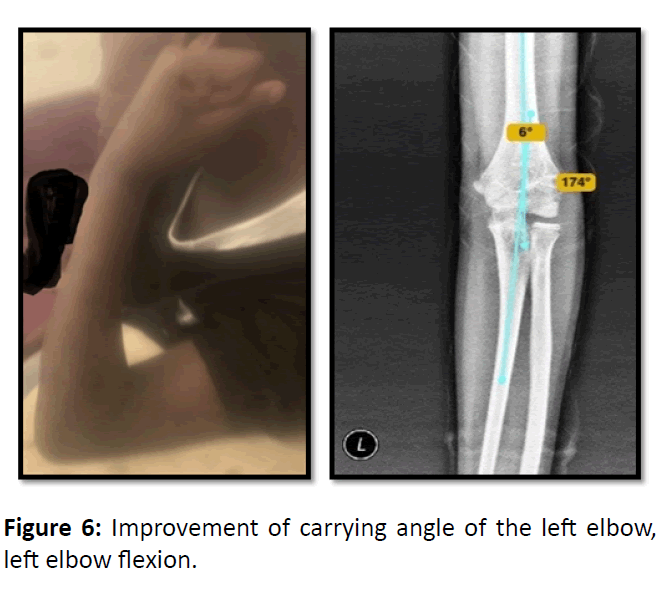
Patient underwent aggressive physical therapy sessions for 4 months, which showed end result as left elbow 120 degree flexion and new x-ray with improvement of carrying angle (Figure 6).
Discussion
Cubits varus deformity is one of the most common complication that seen in children following trauma [3]. Most of the literature supports that malreduction is the main leading cause to this deformity [4]. Indication of Treatment in cubitus varus primarily for cosmetic reasons as majority of the patients is asymptomatic with no functional impairment [5]. Various surgical procedures including corrective osteotomies have been described for correction of this deformity; each procedure has its advantages, disadvantages and possibility of loss of correction [6]. In the presented case, improvement of degree of deformity and carrying angle were achieved both clinically and radiologicaly with minimal complication.
Conclusion
Hemiepiphysiodesis of the lateral condyle is effective method in correcting cubitus varus deformity both clinically and radiologically with safe and easy technique but need for 2nd surgery for implant removal and close follow up.
References
- Patwardhan S, Shyam AK (2015) Cubitus varus deformity–Rationale of treatment and methods. Int J Paediatr Orthop 1: 26-29.
- Marquis CP, Cheung G, Dwyer JS, Emery DF (2008) Supracondylar fractures of the humerus. Cur Orthop 22: 62-69.
- Sashaank SS, Giriraj JK, Menon PG (2017) Deformity correction in cubitus varus-Our experience. IOSR J Dent Med Sci 16: 132-137.
- Verka PS, Kejariwal U, Singh B (2017) Management of cubitus varus deformity in children by closed dome osteotomy. J Clin Diagn Res 11: RC08.
- Dhar SA, Dar TA, Mir NA (2017) Problems, difficulties, and surgical complications of cubitus varus. Curr Orthop Pract 28: 84-88.
- Jain AK, Dhammi IK, Arora A, Singh MP, Luthra JS (2000) Cubitus varus: Problem and solutions. Arch Orthop Traum Su 120: 420-425.

Open Access Journals
- Aquaculture & Veterinary Science
- Chemistry & Chemical Sciences
- Clinical Sciences
- Engineering
- General Science
- Genetics & Molecular Biology
- Health Care & Nursing
- Immunology & Microbiology
- Materials Science
- Mathematics & Physics
- Medical Sciences
- Neurology & Psychiatry
- Oncology & Cancer Science
- Pharmaceutical Sciences
