CARDIOLOGIST
Nasih Ahmed
Nasih Ahmed*
Consultant Cardiologist at Hawler Private Cardiac Hospital, Iraq
*Corresponding author: Nasih Ahmed, Consultant Cardiologist at Hawler Private Cardiac Hospital, Iraq; Tel: 7504531509 ; E-Mail: nasihmohsin@gmail.com
Received date: October 07, 2020, 2020; Accepted date: August 19, 2021; Published date: August 30, 2021
Citation: Nasih A (2021) cardiologist. Med Case Rep Vol.7:8
Abstract
H.cyst rarely involves the heart although in most cases LV is involved but other areas of theheart also could be involved, presentations may differ and it depends on localization of the cyst, size and complications. In this article we reviewed 3 cases of cardiac H.Cyst which have different presentations and different localizations in the heart.
Keywords: Hydatid cyst, Multislice Ct, Electrocardiography, MRI, Right ventricle, Left ventricle.
Introduction
Hydatid cyst is a parasitic human infection more seen in sheep- farming areas. It is caused by Echinococcus granulosis. Humans can be infected by ingesting tapeworm eggs, from which cysts will be developed mostly in the liver and the lung (1) it can affect any organs in the body. They are most common in the liver (50%–70%) and the lung (20–30%) (2-3). Cardiac involvement in H cyst disease is rare, constituting only 0.5–2% of all cases of hydatid disease, Areas of cardiac involvement in H.cyst disease include the left ventricle (60% ), right ventricle (10%),pericardium (7%),pulmonary artery(6%),and left atrial appendage (6%).involvement of the interventricular septum is rare (4% of cases) (4) presentations depend on size and location of the cyst, symptoms are different which might be asymptomatic or present with chest pain. Shortness of breath or might present with complications of ruptured H.cyst (5).
Imaging technique
All patients are informed and written consent taken from them. All patients underwent ECG. Echocardiography and ECG gated cardiac CT scan with and without contrast. All patients underwent contrast CT cardiac angiography examination by using ECG gated cardiac CT ( ICT,philips Medical System). Non contrast study was performed for all patients prior to contrast study. Renal function test assessed for all patient prior to the contrast study. Intravenous access was obtained .65-70 ml of contrast containing 350 mg of Iodine/ml was injected at a flow rate of 5-6 ml/sec then 40-50 ml of normal saline was injected. The data reconstructed at 40% and 78% of the RR interval and transferred to a workstation for analysis.
In this article we reviewed 3 different cases of cardiac H.cyst which has different localization in the heart and different presentations.
Case No. 1:
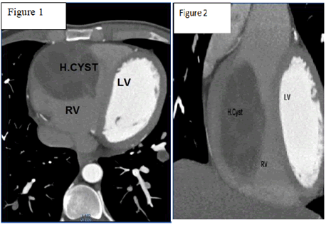
47 years old male patient presented with exertional shortness of breath. ECG was normal. The echocardiography study revealed enlarged RV size and round intracavitory mass in RV with moderate tricuspid regurgitation the patient referred for cardiac CT for further evaluation. The CT images revealed large multilocular non enhancing cystic mass inside the mid RV measuring 70x50mm in size (figure 1&2).decision made for operation the cyst removed succefully without complications. The operative finding was consistent with the H.cyst.
Case No 2:
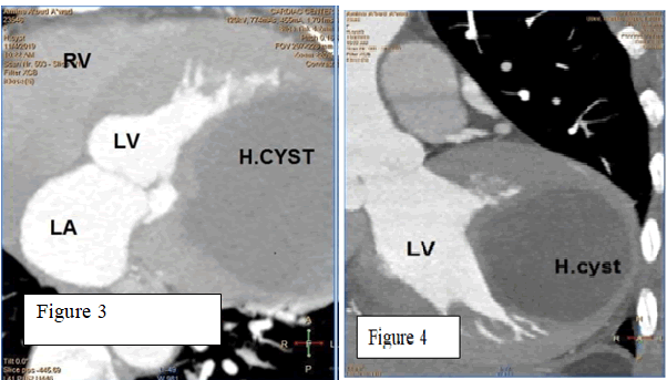
55 years old female patient presented with chest pain. An ECG finding was T wave inversion in V3.V4.V5. echocardiographic study ravelled mass in LV. Cardiac CT angiography revealed Large non enhancing unilocular cystic mass localized to the lateral LV myocardial wall measuring (72mmx65mm) in caliber the patient underwent operation and the cyst removed successfully. The operative finding was consistent with the H.cyst.
Case No 3:
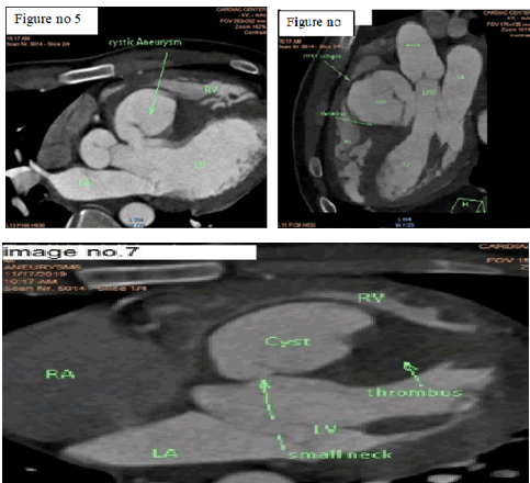
22 years old male patient presented with left side weakness and attacks of shortness of breath, brain MRI revealed ischemic stroke the patient had history of Operation for H.cyst in the liver before 2 years the patient referred for echocardiography examination to search for the underlying cardiac cause of the stroke. The echocardiographic study showed large aneurysmal pouch localized to the base of IVS wall and protruding to RV& RVOT, the mass was connected though a small neck to the LV cavity and the cyst lumen was partially filled by the thrombus. The patient referred for Cardiac CTA for further evaluation cardiac CT revealed large aneurysmal cystic mass localized to the basal portion of IVS wall and connected through small neck to LV cavity and the cystic cavity was partially filled by thrombus and out pouching toward the RV causing significant RVOT narrowing. A serological marker was positive for H.cyst the patient treated conservatively.
Discussion
Hydatid disease is endemic in Mediterranean countries (5) cardiac involvement is rare. The left ventricle is the commonest site of involvement by H.cysts (6), cardiac H.cysts is usually Asymptomatic and only 10% of patients may present with symptoms (7). In first case the patient presented with Dysponea the symptom most likely related to the large size of the cyst which occupying most of the RV cavity and causing RV overload with absence of other underlying causes that could be related to his dysponea which has been excluded simultaneously by CT angiography like pulmonary embolism and other parenchymal lung diseases. Findings of Contrast Cardiac CT was highly suggestive of H.cyst in this patient and positive serological test confirmed the diagnosis
In the second case the patient presented with chest pain because Cardiac H.cysts sometimes with intra-cavity expansion, result in local ischemia to myocardium by pressure effect and sometimes eroding into adjacent areas (8). The findings of CT angiography were highly suggestive for H.cyst and confirmed with positive serological test for Hydatid disease. The first 2 cases underwent open heart surgery cardiopulmonary bypass was used after quilting the cystic cavities all cysts were cleaned and daughter cysts were removed .The cavities were closed. Mebendazole was administered postoperatively. Cardiac hydatid cysts may cause serious complications including cyst rupture anaphylactic shock ,pericardial tamponade .pulmonary embolization, cerebral or peripheral arterial embolism (9) cardiac Hydatid cyst is a rare cause of embolization (10) but it can be a presenting symptom as its seen in the third case in which rupture of the H.cyst resulted cerebral embolization and stroke. Right sided cardiac hydatid cyst have more tendency to expand inside the RV cavity, whereas left-sided H.cysts tends to grow sub epicardially (3) and this is evident in our case reports.
Hydatid cyst of the interventricular septum is rare but it can cause serious complications(9) and this is evident in clinical history of third case report in which the patient developed stroke after rupture of the H.cyst in Interventricular septum. Diagnosis of H.cyst depends on the clinical history. Serologic test and imaging findings. ELISA test is one of the most specific tests that can be used for diagnosis and a positive result confirms the diagnosis (11). Echocardiography provides important findings on size, number of cysts, location’s and relationships with adjacent structures (12) CT or MRI is used for define the localization and morphologic features of hydatid cysts. Calcification, presence of daughter cysts, and detachment of the membrane are specific signs for identification. calcification is best seen on CT but structure of the cyst and its relation with surrounding structure are best seen with MRI (3) in the diagnosis of these 3 case reports we depended on Cardiac CT angiography and positive serological tests .we preferred ECG gated cardiac CT for examination as there is less artifacts from cardiac motion and respiratory artifacts.
MRI scan was not easily feasible for our patients and it has been not done. Surgery is the treatment of choice for management of cardiac H.cyst. Although anthelminthic drugs have been used in the preoperative and postoperative periods, but surgical removal of the cyst is recommended (4,13).
Conclusion
Cardiac H.Cyst is rare and patients may be presented with atypical symptoms or may be asymptomatic and some patients may have serious complications. The clinical scenario of our case reports reveals the importance of early diagnosis and treatment of cardiac H.cyst and also the importance MSCT in the diagnosis as well as it is role in surgical planning and management.
References
- Chadly A M D, Krimi (2004) Cardiac Hydatid Cyst Rupture as Cause of Death. The American
- Journal of Forensic Medicine and Pathology. 25:3 262-264.
- Eckert J, Deplazes P (2004) Biological, epidemiological, and clinical aspects of echinococcosis,
- a zoonosis of increasing concern. Clin Microbiol Rev 17:107-135.
- Memduh Dursun, Ege Terzibasioglu, Cardiac Hydatid Disease: CT and MRI Findings.
- AJR:190, January 2008.
- Onursal E, Elmaci TT, Tireli E, Dindar A, Atilgan D, Ozcan M et al. (2001) Surgical treatment of
- cardiac echinococcosis: report of eight cases. Surg 31:325â??330.
- Johannes Eckert and Peter Deplazes (2004) Biological, Epidemiological, and Clinical Aspects of
- Echinococcosis, a Zoonosis of Increasing Concern, CLINICAL MICROBIOLOGY REVIEWS, 107â??135.
- Fennira, Kamoun B (2019) Cardiac hydatid cyst in the interventricular septum: A literature
- review/ International Journal of Infectious Diseases. 88:120â??126.
- Niarchos C, Kounis GN, Frangides CR, Koutsojannis CM, Batsolaki M, GouvelouDeligianni GV, Kounis NG et al. (2007) Large hydatic cyst of the left ventricle associated with syncopal attacks. Int J Cardiol. 118(1):24-26.
- Ben-Hamda K, Maatouk F, Ben-Farhat M, Betbout F, Gamra H, Addad F, et al. (2003) Eighteenyear experience with echinococcosus of the heart: clinical and echocardiographic features
- in 14 patients. Int J Cardiol. 91(2-3):145-51.
- Kaplan M, Demirtas M, Cimen S, Ozler A (2001) Cardiac hydatid cysts with intracavitary
- Ann Thorac Surg. 71(5):1587-1590.
- Eskild Petersen, Aarhus, Denmark (2019) A large cardiac hydatid cyst in the interventricular
- septum: A case report. International Journal of Infectious Diseases. 78:31â??33.
- Ozer N (2001) Hydatid cyst of the heart as a rare cause of embolization: report of 5 cases
- and review of published reports. J Am Soc Echocardiogr. 14:299â??302.
- Sagl ñcan Y, Yalçñn O, Kaygusuz E (2016) Cystic Echinococcosis: one entity, two unusual
- Turkiye Parazitol Derg. 40:51â??3.

Open Access Journals
- Aquaculture & Veterinary Science
- Chemistry & Chemical Sciences
- Clinical Sciences
- Engineering
- General Science
- Genetics & Molecular Biology
- Health Care & Nursing
- Immunology & Microbiology
- Materials Science
- Mathematics & Physics
- Medical Sciences
- Neurology & Psychiatry
- Oncology & Cancer Science
- Pharmaceutical Sciences


