Keywords
|
| Extra-mammary perianal Paget's disease (EPPD); Multiple tubulovillous adenomas; Colonoscopy |
Introduction
|
| Extra-mammary perianal Paget's disease (EPPD) is a rare skin disorder of unknown aetiology, which is frequently associated with malignancy. Here we report a case of recurrent perianal Paget’s disease which has no association with a malignancy and comments upon its diagnosis and treatment. |
Case
|
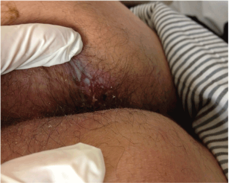
| A 58 year-old man was referred to our dermatology department because of a perianal skin lesion that was recognized during a colonoscopy procedure which was performed for constipation. His lesion was a 4 cm round plaque which was erythematous at the center with a surrounding rim of whitish membranous appearance in the right perianal area (Figure 1). He had pruritus on that area for years. The patient told that he had a similar condition which was operated two years ago. |
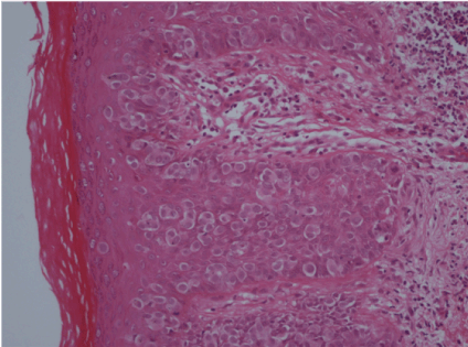
| Histopathologic examination of his skin biopsy was reported as epidermal infiltration of polygonal atypical cells with round pleomorphic nucleus and prominent nucleoli and wide pale eosinophylic cytoplasm along the basal membrane (Figure 2). |
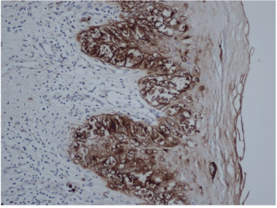
| These atypical cells were heavily stained by carsinoembryogenic antigene (CEA) immunohistochemically (Figure 3). No other colonic, intestinal, or urogenital mass or sign of neoplasm was detected by imaging modalities. It was revealed that he had undergone the similar processes 2 years ago. The patient is diagnosed as primary recurrent perianal Paget’s disease (PPD). An operation for a wide local excision was planned. |
Discussion
|
| Extramammary Paget's disease can be found in various locations, including the patients typically complain of anal pruritus, bleeding, and discharge [1]. Approximately 12% of patients with anal margin Paget's disease harbor a colorectal neoplasm mandating full colonoscopy and 24% of patients have associating cutaneous adnexal adenocarsinoma [2]. |
| No malignancy was detected in our patient by abdominal ultrasound and computed tomography, a simple polyp was excised during colonoscopy in the rectum, which was reported as fibroid polyp histopathologically. |
| Pruritus ani was the main complaint of this patient which was present for many years. The patient had the same story 3 years ago which was ended by excision of the lesion on the right perianal area. Multiple tubulovillous adenomas had been resected during work up at that time. Most authors recommend surgery as the treatment of choice [2-4]. Besa et al. recommends radiotherapy for patients not suitable for surgery, or post operatively [5,6]. Amin reported a successfully treated patient with radiotherapy who had a history as reccurring more extensively four times after each excision [7]. A wide excision is planned for our patient. |
| The differential diagnosis of pruritus ani includes cutaneous candidiasis, tinea cruris, contact dermatitis, intertrigo, lichen simplex chronicus, psoriasis, Bowen disease and even basal cell carcinoma; most of which are seen commonly. PPD is a rare disorder, patients should be examined carefully not to miss the diagnosis. Wide local excision remains the treatment of choice with an evolving role for various adjuvant therapies. Despite adequate resection long term outlook remains guarded due to high recurrence rate. |
Figures at a glance
|
 |
 |
 |
| Figure 1 |
Figure 2 |
Figure 3 |
|
References
|
- Daniel Leonard, David Beddy, Eric JDozois (2011) Neoplasms of Anal Canal and Perianal Skin. Clin Colon Rectal Surg24: 54-63.
- Chanda JJ (1985)Extramammary Paget’s disease: prognosis and relationship to internal malignancy. J Am AcadDermatol13:1000-1014.
- Coldiron BM, Goldsmith BA, Robinson JK (1991)Surgical treatment of extramammary Paget’s disease. Cancer 16:387-403.
- Lee SC, Roth LM, Ehrilch C, Hall JA (1997)Extramammary Paget’s disease of the vulva: a clinicopathologic study of 13 cases. Cancer 39:2540-2549.
- Shutze WP, Gleysteen JJ (1990) Perianal Paget’s disease, classification and management. Report of two cases. Dis Colon Rectum 33:502-507.
- Besa P, Rich TA, Delclos L, Edwards CL, Ota DM, Wharton JT (1992)Extramammary Paget’s disease of the perianal skin. Int J RadiatOncolBiolPhys 24: 73-78.
- Amin R (1999) Perianal Paget’s disease. The British Journal of Radiology 72:610-612.
|


